Acid-beta-glucosidase
From Proteopedia
(Difference between revisions)
| Line 40: | Line 40: | ||
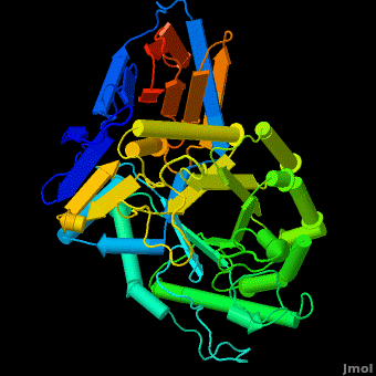
Acid-beta-glucosidase for treatment of a Gaucher disease is produced in CHO cells (<scene name='Acid-beta-glucosidase/Overlay/6'>Cerezyme</scene>™) ([[1ogs]];[[1y7v]];[[2f61]]; [[2nt0]]; [[2nt1]]; [[2v3d]]; [[2v3e]]; [[2nsx]]; [[2j25]]) or in a carrot stem cells suspension (plant produced enzyme by Protalix biopharmaceuticals, [[2v3d]]; [[2v3f]]; [[2v3e]]). Recently solved structures of both enzymes indicate the enzymes are virtually <scene name='Acid-beta-glucosidase/Overlay/7'>identical</scene> in their 3D structures. Yet some small insignificant differences in sugars arise due to the use of different expression systems. | Acid-beta-glucosidase for treatment of a Gaucher disease is produced in CHO cells (<scene name='Acid-beta-glucosidase/Overlay/6'>Cerezyme</scene>™) ([[1ogs]];[[1y7v]];[[2f61]]; [[2nt0]]; [[2nt1]]; [[2v3d]]; [[2v3e]]; [[2nsx]]; [[2j25]]) or in a carrot stem cells suspension (plant produced enzyme by Protalix biopharmaceuticals, [[2v3d]]; [[2v3f]]; [[2v3e]]). Recently solved structures of both enzymes indicate the enzymes are virtually <scene name='Acid-beta-glucosidase/Overlay/7'>identical</scene> in their 3D structures. Yet some small insignificant differences in sugars arise due to the use of different expression systems. | ||
| + | |||
| + | == 3D structures of Acid-β-glucosidase == | ||
| + | [[Acid-β-glucosidase 3D structures]] | ||
| + | |||
</StructureSection> | </StructureSection> | ||
== 3D structures of Acid-β-glucosidase == | == 3D structures of Acid-β-glucosidase == | ||
| Line 50: | Line 54: | ||
[[2nsx]] – hABG + pharmacological chaperone<br /> | [[2nsx]] – hABG + pharmacological chaperone<br /> | ||
[[2xwd]], [[2xwe]] – hABG + nojirimycin derivative<br /> | [[2xwd]], [[2xwe]] – hABG + nojirimycin derivative<br /> | ||
| - | + | ||
== References == | == References == | ||
Revision as of 11:07, 26 February 2019
| |||||||||||
3D structures of Acid-β-glucosidase
Updated on 26-February-2019
2wkl, 3gxd, 3gxi, 3gxm, 2v3f, 2nt0, 2nt1, 2j25, 2f61, 1ogs, 3rik – hABG – human
3ke0, 3keh – hABG (mutant)
2wcg, 3gxf, 2vt0, 2v3e, 2v3d, 1y7v, 3ril, 5lvx – hABG + inhibitor
2nsx – hABG + pharmacological chaperone
2xwd, 2xwe – hABG + nojirimycin derivative
References
- ↑ Dvir H, Harel M, McCarthy AA, Toker L, Silman I, Futerman AH, Sussman JL. X-ray structure of human acid-beta-glucosidase, the defective enzyme in Gaucher disease. EMBO Rep. 2003 Jul;4(7):704-9. PMID:12792654 doi:10.1038/sj.embor.embor873
- ↑ Grabowski GA. Gaucher disease and other storage disorders. Hematology Am Soc Hematol Educ Program. 2012;2012:13-8. doi:, 10.1182/asheducation-2012.1.13. PMID:23233555 doi:http://dx.doi.org/10.1182/asheducation-2012.1.13
Additional Resources
Metabolic Disorders
Carbohydrate Metabolism
Treatment of Gaucher disease
Proteopedia Page Contributors and Editors (what is this?)
Michal Harel, Boris Brumshtein, Alexander Berchansky, Joel L. Sussman, Eran Hodis, David Canner
