User:Vinícius M. Neto/Sandbox 1
From Proteopedia
(Difference between revisions)
| Line 20: | Line 20: | ||
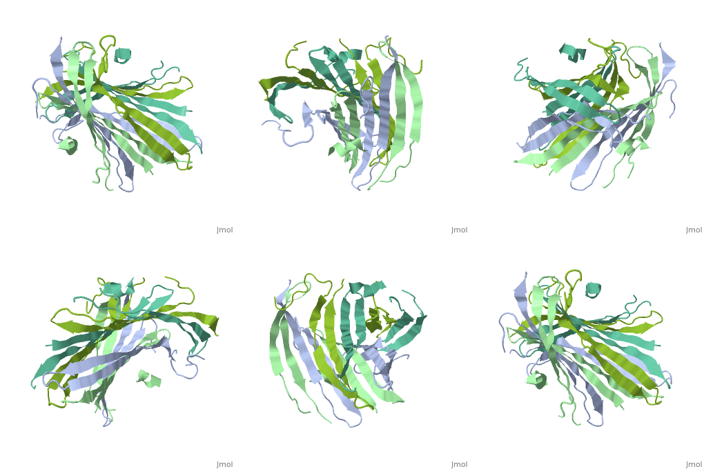
The structure of fibroin is highly pH-dependent. During the natural silk-spinning process, the fibroin solution experiences a steep pH gradient along the silk gland (from anterior to posterior), which triggers the gelation of condensed fibroin. Specifically, the N-terminal domain (FibNT) remains in a disordered random-coil state at neutral pH, preventing premature β-sheet formation. Only when the pH drops to approximately 6.0 does FibNT undergo a cooperative structural transition, adopting the stable β-sheet conformation essential for fiber assembly. | The structure of fibroin is highly pH-dependent. During the natural silk-spinning process, the fibroin solution experiences a steep pH gradient along the silk gland (from anterior to posterior), which triggers the gelation of condensed fibroin. Specifically, the N-terminal domain (FibNT) remains in a disordered random-coil state at neutral pH, preventing premature β-sheet formation. Only when the pH drops to approximately 6.0 does FibNT undergo a cooperative structural transition, adopting the stable β-sheet conformation essential for fiber assembly. | ||
| - | Interactions between acidic residues in FibNT are critical for pH-sensitive behavior. Near the transition point (pH ~6.0), some residues exhibit up-shifted pK<sub>a</sub> values, allowing them to remain ionized at neutral pH. This sustained negative charge creates electrostatic repulsion, actively preventing premature folding and β-sheet assembly. For example, at higher pH, key hydrogen | + | Interactions between acidic residues in FibNT are critical for pH-sensitive behavior. Near the transition point (pH ~6.0), some residues exhibit up-shifted pK<sub>a</sub> values, allowing them to remain ionized at neutral pH. This sustained negative charge creates electrostatic repulsion, actively preventing premature folding and β-sheet assembly. For example, at higher pH, <scene name='10/1082417/Essential_h_bonds/1'>key hydrogen bonds</scene>—such as those between <span style="background-color:black; color:yellow;">'''Glu56–Asp44'''</span> and <span style="background-color:black; color:cyan;">'''Asp100–Glu98'''</span>—are disrupted, destabilizing β-sheet conformations until protonation occurs at lower pH. |
== Relevance == | == Relevance == | ||
Revision as of 19:40, 18 June 2025
Your Heading Here (maybe something like 'Structure')
| |||||||||||
References
- ↑ Hanson, R. M., Prilusky, J., Renjian, Z., Nakane, T. and Sussman, J. L. (2013), JSmol and the Next-Generation Web-Based Representation of 3D Molecular Structure as Applied to Proteopedia. Isr. J. Chem., 53:207-216. doi:http://dx.doi.org/10.1002/ijch.201300024
- ↑ Herraez A. Biomolecules in the computer: Jmol to the rescue. Biochem Mol Biol Educ. 2006 Jul;34(4):255-61. doi: 10.1002/bmb.2006.494034042644. PMID:21638687 doi:10.1002/bmb.2006.494034042644

