Sandbox Reserved 1174
From Proteopedia
| This Sandbox is Reserved from Jan 11 through August 12, 2016 for use in the course CH462 Central Metabolism taught by R. Jeremy Johnson at the Butler University, Indianapolis, USA. This reservation includes Sandbox Reserved 1160 through Sandbox Reserved 1184. |
To get started:
More help: Help:Editing |
Contents |
Human Lysophosphatidic Acid Receptor 1
Example text for references: [1] [2]
Lysophosphatidic Acid
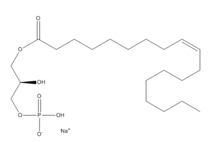
Lysophosphatidic Acid (LPA) consists of an unsaturated fatty acid chain, a glycerol backbone, and a free phosphate group (Figure 1). Lysophosphatidic acid is found in nearly all cells, tissues, and fluids of the body. LPA is present intracellularly as a precursor of phospholipid biosynthesis, and extracellularly as a signalling phospholipid. This page will focus on the signalling role of LPA.
Extracellularly, LPA is produced from lysophosphatidylcholine by the enzyme autotaxin. Autotaxin was originally linked with metastasis, and this link was later discovered to be mediated through the production of LPA, which signals cell proliferation.[3] All of LPA’s activities are receptor mediated; the signalling lipid interacts with at least six G-protein coupled receptors LPA1-LPA6.
Function
Of the six LPA G-protein coupled receptors, LPA1 is the most widely expressed. Lysophosphatidic acid receptor 1 is coupled to a heterotrimeric G protein. The three G alpha proteins that LPA1 couples to are Gi, Gq, and G12/13.[4] From these three G proteins many signal transduction pathways are activated (Figure 2). The downstream effects of Gi are cell proliferation, cell survival, cell migration, and morphological changes. Gq signals the inhibition of gap-junctional communication. Those of G12/13 are morphological changes, inhibition / reversal of differentiation, contraction, and increased endothelial permeability. These downstream functions show the wide array of effects that LPA can have on the body. Targeted deletion of LPA receptors has had an effect on every organ system examined.[5]
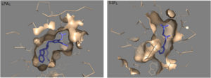
LPA1 is part of the larger EDG (endothelial differentiation gene) family which includes the sphingosine 1-phosphate receptors. Compare to S1P…
Structure
| |||||||||||
Clinical Relevance
Cancer
Fibrosis
Pain
Endocannabinoids
The endocannabinoid system, located in the mammalian nervous system, regulates a variety of physiological processes including appetite, pain sensation, mood, and memory. Endocannabinoids, the natural ligands for cannabinoid receptors, are similar in structure to lysophosphatidic acid. Both the cannabinoid receptors and the LPA receptors have a preference for long unsaturated acyl chains.
References
- ↑ Hanson, R. M., Prilusky, J., Renjian, Z., Nakane, T. and Sussman, J. L. (2013), JSmol and the Next-Generation Web-Based Representation of 3D Molecular Structure as Applied to Proteopedia. Isr. J. Chem., 53:207-216. doi:http://dx.doi.org/10.1002/ijch.201300024
- ↑ Herraez A. Biomolecules in the computer: Jmol to the rescue. Biochem Mol Biol Educ. 2006 Jul;34(4):255-61. doi: 10.1002/bmb.2006.494034042644. PMID:21638687 doi:10.1002/bmb.2006.494034042644
- ↑ Boutin JA, Ferry G. Autotaxin. Cell Mol Life Sci. 2009 Sep;66(18):3009-21. doi: 10.1007/s00018-009-0056-9. Epub , 2009 Jun 9. PMID:19506801 doi:http://dx.doi.org/10.1007/s00018-009-0056-9
- ↑ Moolenaar WH, van Meeteren LA, Giepmans BN. The ins and outs of lysophosphatidic acid signaling. Bioessays. 2004 Aug;26(8):870-81. PMID:15273989 doi:http://dx.doi.org/10.1002/bies.20081
- ↑ 5.0 5.1 5.2 Chrencik JE, Roth CB, Terakado M, Kurata H, Omi R, Kihara Y, Warshaviak D, Nakade S, Asmar-Rovira G, Mileni M, Mizuno H, Griffith MT, Rodgers C, Han GW, Velasquez J, Chun J, Stevens RC, Hanson MA. Crystal Structure of Antagonist Bound Human Lysophosphatidic Acid Receptor 1. Cell. 2015 Jun 18;161(7):1633-43. doi: 10.1016/j.cell.2015.06.002. PMID:26091040 doi:http://dx.doi.org/10.1016/j.cell.2015.06.002


