Image:1742-4690-7-18-7.jpg
From Proteopedia
No higher resolution available.
1742-4690-7-18-7.jpg (600 × 260 pixel, file size: 46 KB, MIME type: image/jpeg)
Summary
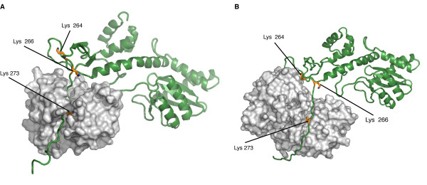
(A) Three-dimensional model of the IN/GCN5 complex. IN is represented in green and GCN5 in light grey. (B) Three-dimensional model of the IN/p300 complex. IN is represented in green and p300 in light grey. In (A) and (B), the three lysine residues in the C-terminal domain of IN that are acetylated by both GCN5 and p300 (Lys 264, Lys 266, and Lys 273) are shown in yellow. GCN5 and p300 are rendered as surfaces, while IN as a cartoon to highlight the C-terminal unfolded portion which inserts in the binding pockets of the two HATs.
Licensing
{{subst:autodate|AutoReplaceable fair use people}}
File history
Click on a date/time to view the file as it appeared at that time.
| Date/Time | User | Dimensions | File size | Comment | |
|---|---|---|---|---|---|
| (current) | 20:54, 5 January 2015 | Charlene Hartnagel (Talk | contribs) | 600×260 | 46 KB | (A) Three-dimensional model of the IN/GCN5 complex. IN is represented in green and GCN5 in light grey. (B) Three-dimensional model of the IN/p300 complex. IN is represented in green and p300 in light grey. In (A) and (B), the three lysine residues in the |
- Edit this file using an external application
See the setup instructions for more information.
Links
The following pages link to this file:
