Image:716px-6VSB spike protein SARS-CoV-2 monomer in homotrimer.png
From Proteopedia
Size of this preview: 398 × 600 pixels
Full resolution (716 × 1079 pixel, file size: 771 KB, MIME type: image/png)
https://commons.wikimedia.org/wiki/File:6VSB_spike_protein_SARS-CoV-2_monomer_in_homotrimer.png#file
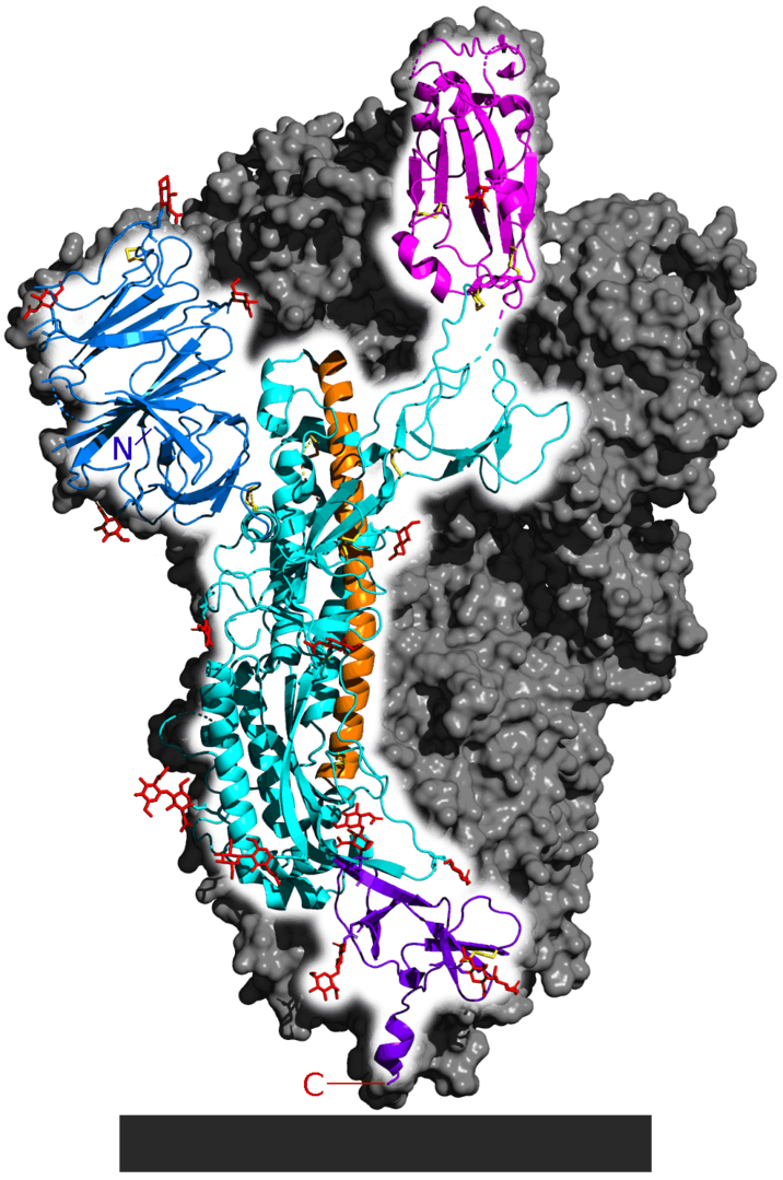
Spike glycoprotein from SARS-CoV-2. PDB: 6VSB. Only one monomer is highlighted. Whole protein is a homotrimer. Rest of the trimer is shown as a gray surface. Parts of the actual structure are not shown. The following are listed from N-terminal (letter N) to C-terminal (C): N-terminal domain (blue), ACE2 receptor binding domain (magenta) general structure (cyan), central helix (orange, faces inside of the homotrimer) and connector domain (purple, anchors the spike protein to virus lipid envelope). Yellow: disulfide bonds. Red: carbohydrates. Gray block: lipid membrane of the virus.
File history
Click on a date/time to view the file as it appeared at that time.
| Date/Time | User | Dimensions | File size | Comment | |
|---|---|---|---|---|---|
| (current) | 20:42, 17 September 2020 | Jeremiah C Hagler (Talk | contribs) | 716×1079 | 771 KB | https://commons.wikimedia.org/wiki/File:6VSB_spike_protein_SARS-CoV-2_monomer_in_homotrimer.png#file Spike glycoprotein from SARS-CoV-2. PDB: 6VSB. Only one monomer is highlighted. Whole protein is a homotrimer. Rest of the trimer is shown as a gray surf |
- Edit this file using an external application
See the setup instructions for more information.
Links
The following pages link to this file:
