Image:Binding pocket try 2.png
From Proteopedia

Size of this preview: 600 × 600 pixels
Full resolution (2000 × 2000 pixel, file size: 1.31 MB, MIME type: image/png)

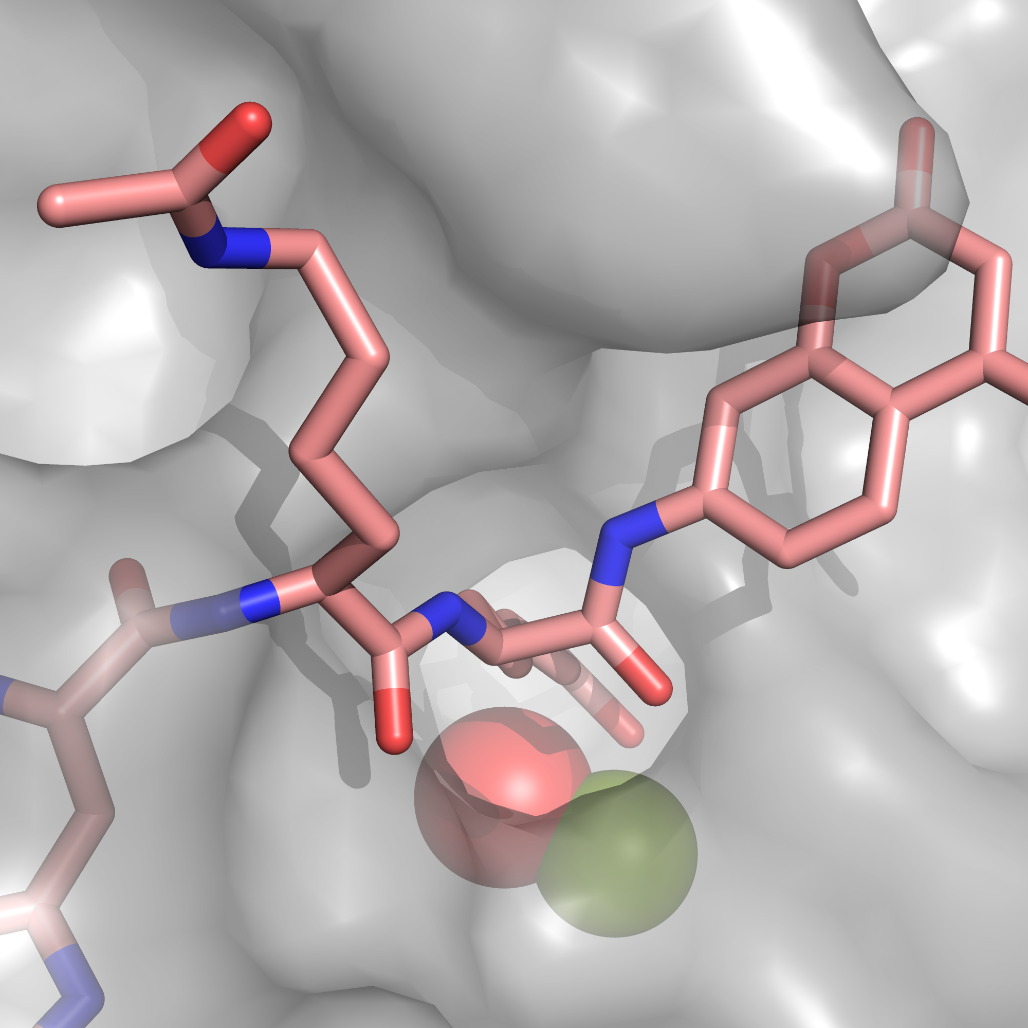
A view inside of the binding pocket of HDAC8, showing the lysine that will be deacetylated.
File history
Click on a date/time to view the file as it appeared at that time.
| Date/Time | User | Dimensions | File size | Comment | |
|---|---|---|---|---|---|
| (current) | 17:43, 22 March 2019 | Carolyn Hurdle (Talk | contribs) | 2000×2000 | 1.31 MB | Reverted to version as of 17:42, 22 March 2019 |
| 17:42, 22 March 2019 | Courtney Brown (Talk | contribs) | 2000×2000 | 1.31 MB | A view of the lysine that will be deacetylated protruding into the binding pocket, with the nucleophilic water and zinc ion visible inside. | |
| 17:42, 22 March 2019 | Carolyn Hurdle (Talk | contribs) | 2000×2000 | 1.31 MB | A view inside of the binding pocket of HDAC8, showing the lysine that will be deacetylated. |
- Edit this file using an external application
See the setup instructions for more information.
Links
There are no pages that link to this file.
