Image:Diels-AlderaseSurfaces.png
From Proteopedia
Size of this preview: 800 × 543 pixels
Full resolution (1079 × 732 pixel, file size: 601 KB, MIME type: image/png)
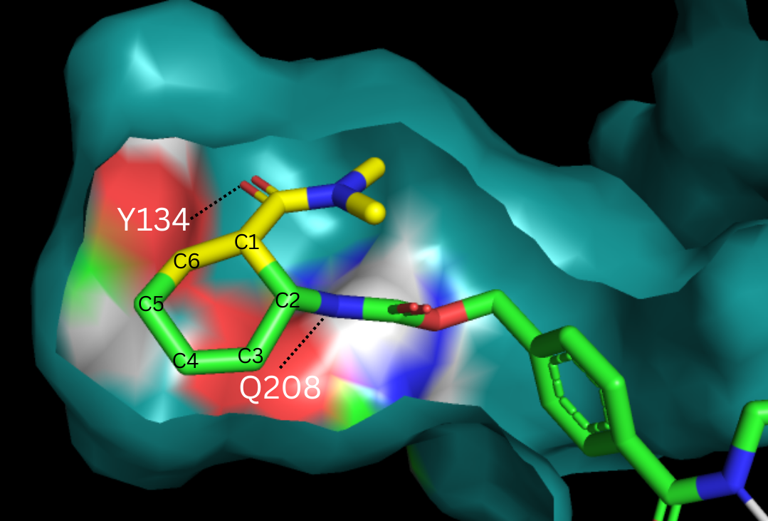
Shown is the binding pocket of the enzyme shown as surface, highlighting the electrostatics of the two catalytic residues, Tyr134 and Glu208. The ligand is color coded based on original structure: the diene is in yellow and the dienophile is in green. The reaction proceeds via attack of the C6 on the C5, shifting electron density to C2, which attacks C1.
File history
Click on a date/time to view the file as it appeared at that time.
| Date/Time | User | Dimensions | File size | Comment | |
|---|---|---|---|---|---|
| (current) | 18:52, 17 April 2025 | Micah Zile (Talk | contribs) | 1079×732 | 601 KB | Shown is the binding pocket of the enzyme shown as surface, highlighting the electrostatics of the two catalytic residues, Tyr134 and Glu208. The ligand is color coded based on original structure: the diene is in yellow and the dienophile is in green. The |
- Edit this file using an external application
See the setup instructions for more information.
Links
The following pages link to this file:
