Image:Figure 3a (Morth et al. 2007).png
From Proteopedia
Size of this preview: 265 × 599 pixels
Full resolution (368 × 832 pixel, file size: 421 KB, MIME type: image/png)
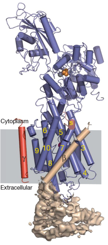
The a-, b- and c-subunits are coloured blue, wheat and red, respectively. Helices are represented by cylinders and b-strands by arrows. The b-ectodomain is shown by surface representation of the experimental electron density. The transmembrane segments of the a-subunit are numbered (yellow) startingwiththemostN-terminal.ThesmallC-terminalhelix(S,forswitch) is light red. Mg21, MgF422 and Rb1 ions are grey, orange and pink, respectively.
File history
Click on a date/time to view the file as it appeared at that time.
| Date/Time | User | Dimensions | File size | Comment | |
|---|---|---|---|---|---|
| (current) | 12:51, 14 July 2020 | Keren Morag (Talk | contribs) | 368×832 | 421 KB | The a-, b- and c-subunits are coloured blue, wheat and red, respectively. Helices are represented by cylinders and b-strands by arrows. The b-ectodomain is shown by surface representation of the experimental electron density. The transmembrane segments o |
- Edit this file using an external application
See the setup instructions for more information.
Links
There are no pages that link to this file.

.png)