Image:General view of beta lactoglobulin.jpg
From Proteopedia
Size of this preview: 557 × 599 pixels
Full resolution (748 × 805 pixel, file size: 69 KB, MIME type: image/jpeg)
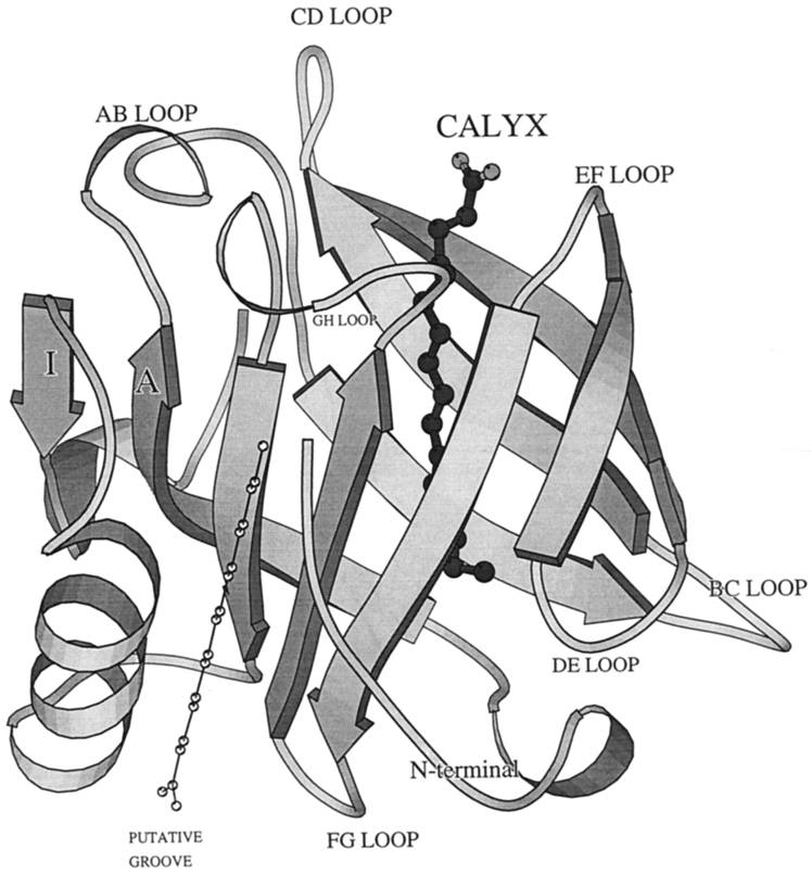
A general view of b-lactoglobulin, a typical lipocalin. The binding site (filled atoms) is shown in the central calyx, and the putative binding site (open atoms) is indicated on the outer surface of the protein. The structurally conserved regions are at the rear of the molecule on strand A, the FG loop, and the loop before the a-helix.(Wu S et al. J. Biol. Chem. 1999;274:170-174)
File history
Click on a date/time to view the file as it appeared at that time.
| Date/Time | User | Dimensions | File size | Comment | |
|---|---|---|---|---|---|
| (current) | 02:49, 7 December 2013 | Rini Triani (Talk | contribs) | 748×805 | 69 KB | A general view of b-lactoglobulin, a typical lipocalin. The binding site (filled atoms) is shown in the central calyx, and the putative binding site (open atoms) is indicated on the outer surface of the protein. The structurally conserved regions are at t |
- Edit this file using an external application
See the setup instructions for more information.
Links
The following pages link to this file:
