Image:Zf rpmodel.jpg
From Proteopedia
No higher resolution available.
Zf_rpmodel.jpg (480 × 360 pixel, file size: 40 KB, MIME type: image/jpeg)
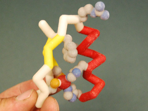
This is a physical model of a zinc finger generated from the PDB file 1ZAA. The model was designed and built by the MSOE Center for BioMolecular modeling using rapid prototyping technology.
In this model, the alpha helix is depicted in red, while the beta sheets are shown in yellow. The residues are shown in CPK format, and the zinc ion bound in the structure is shown in red.
File history
Click on a date/time to view the file as it appeared at that time.
| Date/Time | User | Dimensions | File size | Comment | |
|---|---|---|---|---|---|
| (current) | 21:24, 2 February 2009 | Savannah Anderson (Talk | contribs) | 480×360 | 40 KB | This is a physical model of a zinc finger generated from the PDB file 1ZAA. The model was designed and built by the MSOE Center for BioMolecular modeling using rapid prototyping technology. In this model, the alpha helix is depicted in red, while the be |
- Edit this file using an external application
See the setup instructions for more information.
Links
The following pages link to this file:
Metadata
This file contains additional information, probably added from the digital camera or scanner used to create or digitize it. If the file has been modified from its original state, some details may not fully reflect the modified image.
| Orientation | Normal |
|---|---|
| Horizontal resolution | 72 dpi |
| Vertical resolution | 72 dpi |
| Software used | Adobe Photoshop CS3 Windows |
| File change date and time | 11:03, 24 June 2008 |
| Color space | 65535 |
