User:Wayne Decatur/3fpn Morph methods
From Proteopedia
(Difference between revisions)
(→Morph from normal 3fpn structure to view in Figure 3 of article describing the structure) |
(→Morph from normal 3fpn structure to view in Figure 3 of article describing the structure) |
||
| Line 40: | Line 40: | ||
then save scene in SCENE AUTHORING TOOS with animation that you play once | then save scene in SCENE AUTHORING TOOS with animation that you play once | ||
| + | |||
| + | |||
| + | Then to get SCENE where wireframe on all the time paste in without the 1.8 limit: | ||
| + | select 219:a and sidechain; wireframe 0.55; color cpk; | ||
| + | select 183:b and sidechain; wireframe 0.55;color cpk; | ||
| + | select 170:a and sidechain; wireframe 0.55; color cpk; | ||
| + | select 231:b and sidechain; wireframe 0.55;color cpk; | ||
| + | |||
| + | Save as palindrome! | ||
--> | --> | ||
Revision as of 05:30, 23 October 2009
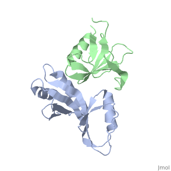
Moving to match Figure 3
Using Pymol and the 3fpn file, I moved so interface is perpendicular to y axis:
translate [10,0,0], chain b
rotate y, 65, chain b
Saved molecule.
Morph from normal 3fpn structure to view in Figure 3 of article describing the structure
|
Took the two files and submitted them. Since the structures didn't have nucleic acids, I took the advice here and used the Yale Morph Server for morphing complexes.
Uploaded to Proteopedia Image:3fpntorotatedversion.pdb.
loaded '3fpntorotatedversion.pdb' in Scene Authoring Tools.
Paper on the structure
- Pakotiprapha D, Liu Y, Verdine GL, Jeruzalmi D. A structural model for the damage-sensing complex in bacterial nucleotide excision repair. J Biol Chem. 2009 May 8;284(19):12837-44. Epub 2009 Mar 13. PMID:19287003 doi:10.1074/jbc.M900571200
