This old version of Proteopedia is provided for student assignments while the new version is undergoing repairs. Content and edits done in this old version of Proteopedia after March 1, 2026 will eventually be lost when it is retired in about June of 2026.
Apply for new accounts at the new Proteopedia. Your logins will work in both the old and new versions.
Sandbox 154
From Proteopedia
| Line 2: | Line 2: | ||
= F-Actin = | = F-Actin = | ||
'''Filamentous actin''' ('''F-actin''') is also referred to as [http://en.wikipedia.org/wiki/microfilaments/ microfilament] <ref> Microfilament - Wikipedia, the free encyclopedia. http://en.wikipedia.org/wiki/Microfilaments. Date accessed: March 16th, 2010. </ref> and is a highly conserved proteinous component found near ubiquitously in eukaryotic cytoskeletons. F-actin and other [http://en.wikipedia.org/wiki/actin/ actin] proteins generally provide a structural role to the cell. | '''Filamentous actin''' ('''F-actin''') is also referred to as [http://en.wikipedia.org/wiki/microfilaments/ microfilament] <ref> Microfilament - Wikipedia, the free encyclopedia. http://en.wikipedia.org/wiki/Microfilaments. Date accessed: March 16th, 2010. </ref> and is a highly conserved proteinous component found near ubiquitously in eukaryotic cytoskeletons. F-actin and other [http://en.wikipedia.org/wiki/actin/ actin] proteins generally provide a structural role to the cell. | ||
| - | + | ||
| - | + | ||
== Introduction == | == Introduction == | ||
| Line 11: | Line 10: | ||
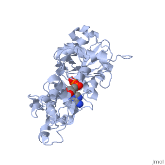
<applet load='2zwh' size='175' color='white' frame='true' align='left' caption='Filamentous Actin (F-actin)'/> | <applet load='2zwh' size='175' color='white' frame='true' align='left' caption='Filamentous Actin (F-actin)'/> | ||
<scene name='Sandbox_154/Thing_in_the_middle/1'>This thing in the middle</scene> | <scene name='Sandbox_154/Thing_in_the_middle/1'>This thing in the middle</scene> | ||
| + | == History of the structure == | ||
| + | The F-actin structure was discovered by Straub in 1942. The structure was speculated based on a low-resolution x-ray crystallograph found in 1990 by Holmes et al. The structure was deposited in the PDB databank in Decemeber 2008 by Oda et al. <ref> Oda T, Iwasa M, Aihara T, Maéda Y, and Narita A. 2009. The nature of the globular-to fibrous actin transition. Nature,457(7228):441-445. PMID: [http://www.ncbi.nlm.nih.gov/pubmed/19158791/ 19158791]</ref>. | ||
Structurally, F-actin appears like a double right-handed helix. It is actually composed of 13 actin units for every 6 left-handed turns, which each have 166° rotations, occurring over 350 Å . <ref> Holmes, K.C., Popp, D., Gebhard, W. and Kabsch, W. 1990. Atomic model of the actin filament. Nature,347(6288):44-49. PMID: [http://www.ncbi.nlm.nih.gov/pubmed/2395461/ 2395461]</ref>. | Structurally, F-actin appears like a double right-handed helix. It is actually composed of 13 actin units for every 6 left-handed turns, which each have 166° rotations, occurring over 350 Å . <ref> Holmes, K.C., Popp, D., Gebhard, W. and Kabsch, W. 1990. Atomic model of the actin filament. Nature,347(6288):44-49. PMID: [http://www.ncbi.nlm.nih.gov/pubmed/2395461/ 2395461]</ref>. | ||
aaaaaaaaaaaaaaaaaaaaaaaaaaaaaaaaaaaaaaaaaaaaaaaaaa | aaaaaaaaaaaaaaaaaaaaaaaaaaaaaaaaaaaaaaaaaaaaaaaaaa | ||
Revision as of 00:49, 26 March 2010
| |||||||
| 2zwh, resolution 3.30Å () | |||||||
|---|---|---|---|---|---|---|---|
| Ligands: | , | ||||||
| Non-Standard Residues: | |||||||
| |||||||
| Resources: | FirstGlance, OCA, RCSB, PDBsum | ||||||
| Coordinates: | save as pdb, mmCIF, xml | ||||||
Contents |
F-Actin
Filamentous actin (F-actin) is also referred to as microfilament [1] and is a highly conserved proteinous component found near ubiquitously in eukaryotic cytoskeletons. F-actin and other actin proteins generally provide a structural role to the cell.
Introduction
Assembly
Structure of F-actin
|
History of the structure
The F-actin structure was discovered by Straub in 1942. The structure was speculated based on a low-resolution x-ray crystallograph found in 1990 by Holmes et al. The structure was deposited in the PDB databank in Decemeber 2008 by Oda et al. [2]. Structurally, F-actin appears like a double right-handed helix. It is actually composed of 13 actin units for every 6 left-handed turns, which each have 166° rotations, occurring over 350 Å . [3]. aaaaaaaaaaaaaaaaaaaaaaaaaaaaaaaaaaaaaaaaaaaaaaaaaa aaaaaaaaaaaaaaaaaaaaaaaaaaaaaaaaaaaaaaaaaaaaaaaaa aaaaaaaaaaaaaaaaaaaaaaaaaaaaaaaaaaaaaaaaaaaaaaaaaaaa aaaaaaaaaaaaaaaaaaaaaaaaaaaaaaaaaaaaaaaaaaaaaaaaaaaaaa aaaaaaaaaaaaaaaaaaaaaaaaaaaaaaaaaaaaaaaaaaaaaaaaaaaaa aaaaaaaaaaaaaaaaaaaaaaaaaaaaaaaaaaaaaaaaaaaaaaaaaa
Monomeric Unit - G-actin
|
aaaaaaaaaaaaaaaaaaaaaaaaaaaaaaaaaaaaaaaaaaaaaaaaaa aaaaaaaaaaaaaaaaaaaaaaaaaaaaaaaaaaaaaaaaaaaaaaaaa aaaaaaaaaaaaaaaaaaaaaaaaaaaaaaaaaaaaaaaaaaaaaaaaaaaa aaaaaaaaaaaaaaaaaaaaaaaaaaaaaaaaaaaaaaaaaaaaaaaaaaaaaa aaaaaaaaaaaaaaaaaaaaaaaaaaaaaaaaaaaaaaaaaaaaaaaaaaaaa aaaaaaaaaaaaaaaaaaaaaaaaaaaaaaaaaaaaaaaaaaaaaaaaaa
Domains
Function
Enzymatic Role
Active Site
Ligand
Structural Role
References
- ↑ Microfilament - Wikipedia, the free encyclopedia. http://en.wikipedia.org/wiki/Microfilaments. Date accessed: March 16th, 2010.
- ↑ Oda T, Iwasa M, Aihara T, Maéda Y, and Narita A. 2009. The nature of the globular-to fibrous actin transition. Nature,457(7228):441-445. PMID: 19158791
- ↑ Holmes, K.C., Popp, D., Gebhard, W. and Kabsch, W. 1990. Atomic model of the actin filament. Nature,347(6288):44-49. PMID: 2395461
| Please do NOT make changes to this Sandbox until after April 23, 2010. Sandboxes 151-200 are reserved until then for use by the Chemistry 307 class at UNBC taught by Prof. Andrea Gorrell. |
