Calcium-free Calmodulin
From Proteopedia
| Line 5: | Line 5: | ||
==='''General Information'''=== | ==='''General Information'''=== | ||
---- | ---- | ||
| - | Calmodulin is a molecule that has been studied extensively in its functions within the cell, and has an important role in relaying Ca2+ signals within the cytosol. <ref name="1CRT">Hoeflich, K.P., & Ikura, M.. Calmodulin in action: diversity in target recognition and activation mechanisms, Cell. 2002 108:739-742</ref> It does this by binding to Ca2+, undergoing a conformational change, and may interact with various proteins within the cell.<ref name= | + | Calmodulin is a molecule that has been studied extensively in its functions within the cell, and has an important role in relaying Ca2+ signals within the cytosol. <ref name="1CRT">Hoeflich, K.P., & Ikura, M.. Calmodulin in action: diversity in target recognition and activation mechanisms, Cell. 2002 108:739-742</ref> It does this by binding to Ca2+, undergoing a conformational change, and may interact with various proteins within the cell.<ref name="1CRT"/><ref name="4CRT">PMID: 7552748</ref><ref name="3CRT">PMID: 3145979</ref> Once bound to a target protein, it undergoes a further conformational change and may activate certain systems. For example, there is a Ca2+ pump in the plasma membrane pump that is activated by the binding of Ca2+-bound calmodulin, and then uses ATP to drive the Ca2+ out of the cell. <ref name="6CRT">PMID: 12838335</ref> |
[[Image:3cln.png|left|200px]] | [[Image:3cln.png|left|200px]] | ||
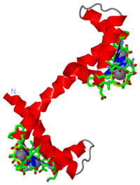
==='''Calcium-bound Calmodulin'''=== | ==='''Calcium-bound Calmodulin'''=== | ||
| - | The structure of calcium-bound calmodulin had previously been discovered using x-ray crystallography <ref name= | + | The structure of calcium-bound calmodulin had previously been discovered using x-ray crystallography <ref name="3CRT"/>. It was then theorized that knowledge of the structure of calcium-free calmodulin would give greater insight into the function of the protein. Attempts were made to crystallize the calcium-free (or apo) form, |
Revision as of 05:45, 1 April 2010
Please do NOT make changes to this Sandbox until after April 23, 2010. Sandboxes 151-200 are reserved until then for use by the Chemistry 307 class at UNBC taught by Prof. [[User:Andrea Gorrell|Andrea Chris Truscott
Calcium-free CalmodulinGeneral InformationCalmodulin is a molecule that has been studied extensively in its functions within the cell, and has an important role in relaying Ca2+ signals within the cytosol. [1] It does this by binding to Ca2+, undergoing a conformational change, and may interact with various proteins within the cell.[1][2][3] Once bound to a target protein, it undergoes a further conformational change and may activate certain systems. For example, there is a Ca2+ pump in the plasma membrane pump that is activated by the binding of Ca2+-bound calmodulin, and then uses ATP to drive the Ca2+ out of the cell. [4] Calcium-bound CalmodulinThe structure of calcium-bound calmodulin had previously been discovered using x-ray crystallography [3]. It was then theorized that knowledge of the structure of calcium-free calmodulin would give greater insight into the function of the protein. Attempts were made to crystallize the calcium-free (or apo) form,
References
|
|||||||||||||||||||||||||||||||||||||||
Proteopedia Page Contributors and Editors (what is this?)
Christopher Truscott, David Canner, Alexander Berchansky, Michal Harel, Andrea Gorrell


