Gyrase
From Proteopedia
| Line 3: | Line 3: | ||
<applet load='3l6v.pdb' size='350' frame='true' align='right' scene="Gyrase/Gyrase_starting_scene/1" caption= 'Crystal Structure of the Xanthomonas campestris Gyrase A C-terminal Domain, [[3l6v]]' /> | <applet load='3l6v.pdb' size='350' frame='true' align='right' scene="Gyrase/Gyrase_starting_scene/1" caption= 'Crystal Structure of the Xanthomonas campestris Gyrase A C-terminal Domain, [[3l6v]]' /> | ||
| - | ''' | + | '''Gyrase (Gyr)''' is a type of topoisomerase II in prokaryotes which unwinds double stranded DNA. The DNA Gyr cutting allows the formation of a negative DNA supercoil which enables replication of DNA. Gyr consists of 2 subunits: GyrA and GyrB. Reverse gyrase (Top-RG) is a type of topoisomerase I which catalyses the formation of positive DNA supercoil. |
| - | + | ||
| - | = | + | {{TOC limit|limit=2}} |
| - | + | ||
| - | + | ||
==3D Structure of Gyrase== | ==3D Structure of Gyrase== | ||
Revision as of 06:15, 26 July 2010
|
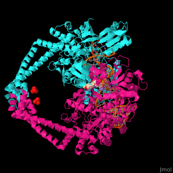
Gyrase (Gyr) is a type of topoisomerase II in prokaryotes which unwinds double stranded DNA. The DNA Gyr cutting allows the formation of a negative DNA supercoil which enables replication of DNA. Gyr consists of 2 subunits: GyrA and GyrB. Reverse gyrase (Top-RG) is a type of topoisomerase I which catalyses the formation of positive DNA supercoil.
3D Structure of Gyrase
Gyrase Subunit A
3l6v – GyrA C-terminal – Xanthomonas campestris
2wl2 – EcGyrA N-terminal+simocylinone – Escherichia coli
1ajb - EcGyrA N-terminal+novobiocin
1zi0, 1ab4 - EcGyrA C-terminal
1x75 – EcGyrA14+CcdB
3ilw - MtGyrA N-terminal – Mycobacterium tuberculosis
1suu - GyrA C-terminal – Borrelia burgdorferi
Gyrase Subunit B
3g75, 3g7b, 3g7e – GyrB+thiazole inhibitor – Staphylococcus aureus
2cjt - MtGyrB C-terminal
3cwv – GyrB truncated – Myxococcus xanthus
1kzn, 1ei1 - EcGyrB N-terminal+clorobiocin
1kij – GyrB domain+novobiocin – Thermus thermophilus
Reverse Gyrase
1gku – AfTop-RG – Archaeoglobus fulgidus
1gl9 - AfTop-RG+ADPNP
Proteopedia Page Contributors and Editors (what is this?)
Michal Harel, Alexander Berchansky, David Canner, Joel L. Sussman
