Journal:PLoS ONE:1
From Proteopedia
(Difference between revisions)

| Line 11: | Line 11: | ||
The hydrophobic pocket is represented by blue spheres and the position of <font color='orange'><b>amino acid 143 of VP1, one of two determinants for neurovirulence in type 2 poliovirus is represented by orange spheres</b></font>. The colors are as in the previous scenes. <scene name='Journal:PLoS_ONE:1/F45/7'>All three substitutions are located adjacent to the 5-fold axis of symmetry</scene>. <scene name='Journal:PLoS_ONE:1/F45/8'>Unique substitutions found in SD-07-03 at residues 141, 144, and 145 of VP1 flank Thr143</scene>. None of SD-0703’s unique substitutions were located near the 3-fold axis of symmetry. | The hydrophobic pocket is represented by blue spheres and the position of <font color='orange'><b>amino acid 143 of VP1, one of two determinants for neurovirulence in type 2 poliovirus is represented by orange spheres</b></font>. The colors are as in the previous scenes. <scene name='Journal:PLoS_ONE:1/F45/7'>All three substitutions are located adjacent to the 5-fold axis of symmetry</scene>. <scene name='Journal:PLoS_ONE:1/F45/8'>Unique substitutions found in SD-07-03 at residues 141, 144, and 145 of VP1 flank Thr143</scene>. None of SD-0703’s unique substitutions were located near the 3-fold axis of symmetry. | ||
| + | <!-- | ||
| + | |||
| + | <scene name='Journal:PLoS_ONE:1/F41/2'>TextToBeDisplayed</scene> | ||
| + | <scene name='Journal:PLoS_ONE:1/F42/1'>TextToBeDisplayed</scene> | ||
| + | <scene name='Journal:PLoS_ONE:1/F51/1'>TextToBeDisplayed</scene> | ||
| + | <scene name='Journal:PLoS_ONE:1/F65/1'>TextToBeDisplayed</scene> | ||
| + | <scene name='Journal:PLoS_ONE:1/F5/1'>TextToBeDisplayed</scene> | ||
| + | <scene name='Journal:PLoS_ONE:1/F55/1'>TextToBeDisplayed</scene> | ||
| + | --> | ||
</StructureSection> | </StructureSection> | ||
| + | |||
<references/> | <references/> | ||
__NOEDITSECTION__ | __NOEDITSECTION__ | ||
Revision as of 07:34, 28 March 2011
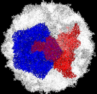
Complete Poliovirus 2 Viron (capsid) based on PDB entry 1eah, example of 3-fold symmetry is in red, example of 5-fold symmetry is in blue.
| |||||||||||
- ↑ DOI
This page complements a publication in scientific journals and is one of the Proteopedia's Interactive 3D Complement pages. For aditional details please see I3DC.

