Sang Joon Won/sandbox 1
From Proteopedia
| Line 12: | Line 12: | ||
==Structure== | ==Structure== | ||
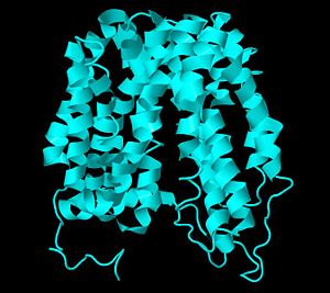
<scene name='Sang_Joon_Won/sandbox_1/Lacy/1'>LacY</scene> has 12 transmembrane helices in which the N- and C-terminal 6 helices form two bundles connected by a loop between helices VI and VII. The 6 helices domains have a pseudo-two fold axis of symmetry and they are positioned to form a large interior <scene name='Sang_Joon_Won/sandbox_1/Lacy/2'>hydrophilic cavity</scene> open on the cytoplasmic side (2). The cavity is composed of helices I, II, IV, and V of the N-terminal domain and helices VII, VIII, X and XI of the C-terminal domain. All the available structures up to date exhibits an inward facing conformation and many studies have indicated that the periplasmic barrier is tightly closed in the absence of sugar binding. Therefore, the inward-facing conformation, which faces the cytoplasm, represents the lowest free-energy state in the membrane (2). A single β-D-galactopyranosyl1-thio-β-D-galactopyranoside (TDG) was bound to the cavity in the X-ray structure to mimic the actual sugar binding and there is only 1 binding site (3). | <scene name='Sang_Joon_Won/sandbox_1/Lacy/1'>LacY</scene> has 12 transmembrane helices in which the N- and C-terminal 6 helices form two bundles connected by a loop between helices VI and VII. The 6 helices domains have a pseudo-two fold axis of symmetry and they are positioned to form a large interior <scene name='Sang_Joon_Won/sandbox_1/Lacy/2'>hydrophilic cavity</scene> open on the cytoplasmic side (2). The cavity is composed of helices I, II, IV, and V of the N-terminal domain and helices VII, VIII, X and XI of the C-terminal domain. All the available structures up to date exhibits an inward facing conformation and many studies have indicated that the periplasmic barrier is tightly closed in the absence of sugar binding. Therefore, the inward-facing conformation, which faces the cytoplasm, represents the lowest free-energy state in the membrane (2). A single β-D-galactopyranosyl1-thio-β-D-galactopyranoside (TDG) was bound to the cavity in the X-ray structure to mimic the actual sugar binding and there is only 1 binding site (3). | ||
| - | |||
Revision as of 16:57, 9 April 2011
Lactose Permease
Background
The lactose permease (LacY) is arguably a paradigm for secondary transporter proteins. This membrane protein belongs to the Major Facilitator Superfamily and has been studied for decades to understand detailed mechanism of energy transduction and translocation reactions. This protein serves as the lactose and hydrogen ion (H+) symporter, utilizing the free energy released from downhill movement of H+ to actively transport lactose. Lactose is a disaccharide that yields D-glucose and D-galactose when hydrolyzed. LacY is specific for disaccharides like lactose that contain a D-galactopyranosyl ring or D- galactose. Due to LacY, higher concentration of sugar molecules can be maintained inside a cell. Keeping higher level of sugar is essential because all bacteria must utilize the energy sources, such as carbohydrate, in their environment in order to produce ATP. ATP provides energy for the biosynthetic processes that bacteria use for their maintenance and reproduction. LacY also uses the energy released from downhill movement of sugar to generate electrochemical H+ gradient [1]. These processes are found in many organisms and play a crucial role in many aspects of cell function. Therefore, many biochemical techniques to study LacY have been applied to the study of many other similar membrane proteins. Since LacY has been used as a model protein for secondary transporter proteins in this era, it is important to understand the structural basis for this transporter because it may give us detailed understanding of energy transduction mechanisms utilized in many living organisms [1].
| |||||||||
| 1pv7, resolution 3.60Å () | |||||||||
|---|---|---|---|---|---|---|---|---|---|
| Ligands: | |||||||||
| Related: | 1pv6 | ||||||||
| |||||||||
| |||||||||
| |||||||||
| Resources: | FirstGlance, OCA, RCSB, PDBsum | ||||||||
| Coordinates: | save as pdb, mmCIF, xml | ||||||||
Structure
has 12 transmembrane helices in which the N- and C-terminal 6 helices form two bundles connected by a loop between helices VI and VII. The 6 helices domains have a pseudo-two fold axis of symmetry and they are positioned to form a large interior open on the cytoplasmic side (2). The cavity is composed of helices I, II, IV, and V of the N-terminal domain and helices VII, VIII, X and XI of the C-terminal domain. All the available structures up to date exhibits an inward facing conformation and many studies have indicated that the periplasmic barrier is tightly closed in the absence of sugar binding. Therefore, the inward-facing conformation, which faces the cytoplasm, represents the lowest free-energy state in the membrane (2). A single β-D-galactopyranosyl1-thio-β-D-galactopyranoside (TDG) was bound to the cavity in the X-ray structure to mimic the actual sugar binding and there is only 1 binding site (3).
- ↑ 1.0 1.1 Guan L, Kaback HR. Lessons from lactose permease. Annu Rev Biophys Biomol Struct. 2006;35:67-91. PMID:16689628 doi:10.1146/annurev.biophys.35.040405.102005


