Group:MUZIC:Telethonin
From Proteopedia

| Line 13: | Line 13: | ||
For the full activation of the gene the regulation of E1 is highly important. MyoD plays an important role in this regulation, and from myogenin in late differentiation of myoblasts. <ref>PMID:21305318</ref> | For the full activation of the gene the regulation of E1 is highly important. MyoD plays an important role in this regulation, and from myogenin in late differentiation of myoblasts. <ref>PMID:21305318</ref> | ||
| - | At the transcriptional level it is thought that there is only one isoform ot Tcap, and it is one of the most abundant transcripts in skeletal muscle <ref>PMID:9350988< | + | At the transcriptional level it is thought that there is only one isoform ot Tcap, and it is one of the most abundant transcripts in skeletal muscle <ref>PMID:9350988</ref>. It does not have different levels of expression in different type of skeletal muscle; levels of expression of Tcap are lower in neonatal compared to adult striated muscle. The transcript is accumulated in a linear pattern similar to that of the myosin heavy chain <ref>PMID: 10208846</ref>. In these same studies it was reported that dennervation lead to decrease in expression of Tcap, suggesting that locomotor activity is also a regulator for its maintainance. |
== T-cap protein == | == T-cap protein == | ||
Telethonin protein is found mostly in skeletal and cardiac muscle. It is one of the major components of the sarcomere, it is localized to the Z-disc. | Telethonin protein is found mostly in skeletal and cardiac muscle. It is one of the major components of the sarcomere, it is localized to the Z-disc. | ||
| - | Studies on telethonin structure by Zou et al. <ref>PMID:16407954< | + | Studies on telethonin structure by Zou et al. <ref>PMID:16407954</ref> report that it is formed of five stranded antiparallel β-sheet extended by two wing-shaped β-hairpin motifs (A-B, C-D LINK). And these two motifs are related by an approximate two-fold symmetry, which generates an almost perfect palindromic arrangement. |
| - | The structure of telethonin was determined using X-ray crystallography <ref>PMID:12446666< | + | The structure of telethonin was determined using X-ray crystallography <ref>PMID:12446666</ref> <ref>PMID16407954</ref> . The shape and architecture of the complex of titin/telethonin was studied by small-angle- X-ray scattering (SAXS) and then compared to the crystallographic models. They also used in-vitro experiments to follow the formation of the complex in non-myogenic Cos1 cells, in order to understand if the assemblage is possible <ref>PMID:16713295</ref> |
This symmetry of telethonin permits its interaction with titin. Both are assembled in an antiparallel sandwich in a (2:1, titin:telethonin). Titin N-terminal domains Z1 and Z2 interact with the N-terminal region of telethonin (residues 1-53). Telethonin mediates in the antiparallel assembly of the two Z1Z2domains. | This symmetry of telethonin permits its interaction with titin. Both are assembled in an antiparallel sandwich in a (2:1, titin:telethonin). Titin N-terminal domains Z1 and Z2 interact with the N-terminal region of telethonin (residues 1-53). Telethonin mediates in the antiparallel assembly of the two Z1Z2domains. | ||
| - | In early differenciating myocyets titin C-terminal and Telethonin co-localize and titin kinase is close to telethonin C-terminal, and it is phosphorilated. This co-localization is not seen in adult myofibrils, titin kinase is reported to localize in the M-band; <ref>PMID:9804419< | + | In early differenciating myocyets titin C-terminal and Telethonin co-localize and titin kinase is close to telethonin C-terminal, and it is phosphorilated. This co-localization is not seen in adult myofibrils, titin kinase is reported to localize in the M-band; <ref>PMID:9804419</ref> It was also informed that telethonin interacts with other proteins including: Potassium channel subuint miK/isk, ankyrin1, and Z-disc proteins FATZ, Calsarcin-3, Ankrd2 <ref>PMID:15136035</ref> and MLP <ref>PMID:12507422</ref>. |
| - | Telethonin is also involved in signalling processes that regulate muscle development. A feed back loop is formed MRFs (MyoD, myogenin, Myf5) regulating Tcap gene expression; telethonin interacts with myostatin inhibiting it. So it regulates MyoD through Myostatin – Smad3 pathway. <ref>PMID:18440815< | + | Telethonin is also involved in signalling processes that regulate muscle development. A feed back loop is formed MRFs (MyoD, myogenin, Myf5) regulating Tcap gene expression; telethonin interacts with myostatin inhibiting it. So it regulates MyoD through Myostatin – Smad3 pathway. <ref>PMID:18440815</ref>. |
| + | == References == | ||
<references/> | <references/> | ||
Revision as of 16:25, 1 July 2011
|
Contents |
Introduction
Also known as T-Cap or Titin Cap protein.
It is a small protein of 19kDa, 167 amino acids. Predominantly expressed in striated muscle. It is a structural protein of the muscle; it is associated to the Z-disc in the sarcomere. It acts as link between titin and other proteins implicated in muscle structure and signalling.
Genetic overview
It is encoded by Tcap gene, in mice (Mus musculus) and humans (Homo sapiens). In mice it is located in chromosome 11, in humans in the long arm of chromosome 17. Tcap is encoded by two exons, and has non-conserved intragenic sequences. The gene is flanked by Stard3 upstream separated by 2,8kb, and Pnmt1 downstream separated by 1,7kb. It has three conserved E-box elements at -103bp (E1), -272bp (E2), and -2067bp (E3). For the full activation of the gene the regulation of E1 is highly important. MyoD plays an important role in this regulation, and from myogenin in late differentiation of myoblasts. [1]
At the transcriptional level it is thought that there is only one isoform ot Tcap, and it is one of the most abundant transcripts in skeletal muscle [2]. It does not have different levels of expression in different type of skeletal muscle; levels of expression of Tcap are lower in neonatal compared to adult striated muscle. The transcript is accumulated in a linear pattern similar to that of the myosin heavy chain [3]. In these same studies it was reported that dennervation lead to decrease in expression of Tcap, suggesting that locomotor activity is also a regulator for its maintainance.
T-cap protein
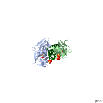
Telethonin protein is found mostly in skeletal and cardiac muscle. It is one of the major components of the sarcomere, it is localized to the Z-disc. Studies on telethonin structure by Zou et al. [4] report that it is formed of five stranded antiparallel β-sheet extended by two wing-shaped β-hairpin motifs (A-B, C-D LINK). And these two motifs are related by an approximate two-fold symmetry, which generates an almost perfect palindromic arrangement.
The structure of telethonin was determined using X-ray crystallography [5] [6] . The shape and architecture of the complex of titin/telethonin was studied by small-angle- X-ray scattering (SAXS) and then compared to the crystallographic models. They also used in-vitro experiments to follow the formation of the complex in non-myogenic Cos1 cells, in order to understand if the assemblage is possible [7]
This symmetry of telethonin permits its interaction with titin. Both are assembled in an antiparallel sandwich in a (2:1, titin:telethonin). Titin N-terminal domains Z1 and Z2 interact with the N-terminal region of telethonin (residues 1-53). Telethonin mediates in the antiparallel assembly of the two Z1Z2domains.
In early differenciating myocyets titin C-terminal and Telethonin co-localize and titin kinase is close to telethonin C-terminal, and it is phosphorilated. This co-localization is not seen in adult myofibrils, titin kinase is reported to localize in the M-band; [8] It was also informed that telethonin interacts with other proteins including: Potassium channel subuint miK/isk, ankyrin1, and Z-disc proteins FATZ, Calsarcin-3, Ankrd2 [9] and MLP [10].
Telethonin is also involved in signalling processes that regulate muscle development. A feed back loop is formed MRFs (MyoD, myogenin, Myf5) regulating Tcap gene expression; telethonin interacts with myostatin inhibiting it. So it regulates MyoD through Myostatin – Smad3 pathway. [11].
References
- ↑ Zhang S, Londhe P, Zhang M, Davie JK. Transcriptional analysis of the titin cap gene. Mol Genet Genomics. 2011 Mar;285(3):261-72. Epub 2011 Feb 9. PMID:21305318 doi:10.1007/s00438-011-0603-6
- ↑ Valle G, Faulkner G, De Antoni A, Pacchioni B, Pallavicini A, Pandolfo D, Tiso N, Toppo S, Trevisan S, Lanfranchi G. Telethonin, a novel sarcomeric protein of heart and skeletal muscle. FEBS Lett. 1997 Sep 29;415(2):163-8. PMID:9350988
- ↑ Mason P, Bayol S, Loughna PT. The novel sarcomeric protein telethonin exhibits developmental and functional regulation. Biochem Biophys Res Commun. 1999 Apr 21;257(3):699-703. PMID:10208846 doi:10.1006/bbrc.1999.0531
- ↑ Zou P, Pinotsis N, Lange S, Song YH, Popov A, Mavridis I, Mayans OM, Gautel M, Wilmanns M. Palindromic assembly of the giant muscle protein titin in the sarcomeric Z-disk. Nature. 2006 Jan 12;439(7073):229-33. PMID:16407954 doi:10.1038/nature04343
- ↑ Zou P, Gautel M, Geerlof A, Wilmanns M, Koch MH, Svergun DI. Solution scattering suggests cross-linking function of telethonin in the complex with titin. J Biol Chem. 2003 Jan 24;278(4):2636-44. Epub 2002 Nov 20. PMID:12446666 doi:10.1074/jbc.M210217200
- ↑ . PMID:216315890657
- ↑ Pinotsis N, Petoukhov M, Lange S, Svergun D, Zou P, Gautel M, Wilmanns M. Evidence for a dimeric assembly of two titin/telethonin complexes induced by the telethonin C-terminus. J Struct Biol. 2006 Aug;155(2):239-50. Epub 2006 Apr 27. PMID:16713295 doi:10.1016/j.jsb.2006.03.028
- ↑ Mayans O, van der Ven PF, Wilm M, Mues A, Young P, Furst DO, Wilmanns M, Gautel M. Structural basis for activation of the titin kinase domain during myofibrillogenesis. Nature. 1998 Oct 29;395(6705):863-9. PMID:9804419 doi:10.1038/27603
- ↑ Kojic S, Medeot E, Guccione E, Krmac H, Zara I, Martinelli V, Valle G, Faulkner G. The Ankrd2 protein, a link between the sarcomere and the nucleus in skeletal muscle. J Mol Biol. 2004 May 28;339(2):313-25. PMID:15136035 doi:10.1016/j.jmb.2004.03.071
- ↑ Knoll R, Hoshijima M, Hoffman HM, Person V, Lorenzen-Schmidt I, Bang ML, Hayashi T, Shiga N, Yasukawa H, Schaper W, McKenna W, Yokoyama M, Schork NJ, Omens JH, McCulloch AD, Kimura A, Gregorio CC, Poller W, Schaper J, Schultheiss HP, Chien KR. The cardiac mechanical stretch sensor machinery involves a Z disc complex that is defective in a subset of human dilated cardiomyopathy. Cell. 2002 Dec 27;111(7):943-55. PMID:12507422
- ↑ Markert CD, Ning J, Staley JT, Heinzke L, Childers CK, Ferreira JA, Brown M, Stoker A, Okamura C, Childers MK. TCAP knockdown by RNA interference inhibits myoblast differentiation in cultured skeletal muscle cells. Neuromuscul Disord. 2008 May;18(5):413-22. Epub 2008 Apr 28. PMID:18440815 doi:10.1016/j.nmd.2008.03.010
