User:Jamie Abbott/Sandbox2
From Proteopedia
| Line 8: | Line 8: | ||
'''Adenosine Triphosphate Binding''' | '''Adenosine Triphosphate Binding''' | ||
| - | Many interactions are required to prepare ATP for attack by a bound histidine molecule and encourage the magnesium pyrophosphate moiety to act as a leaving group. Residues in the β strands and the loop portion of motif 2 are important in ATP contacts for HisRS<ref name="Arnez97">PMID: 9207058</ref>. Generally, residues involved in ATP binding are among the most highly conserved in the HisRS family and for the most part shared by all members in class II. The π-stacking interaction between the adenine ring of ATP and Phe125 provides specificity in the binding of ATP. The recognition of the N6 amino group of ATP involves the main chain carbonyl of Tyr122. The ATP ribose 2’ OH forms an additional contact with HisRS by hydrogen bonding with the main chain carbonyl of Thr281. Furthermore, conserved residues Arg113 and Glu115 stabilize the triphosphate group of ATP in a position such that it points back towards the adenine base. This “fishhook” conformation of ATP is evidently unique to class II aaRS<ref name="Arnez97-2">PMID: 9204708</ref>. | + | Many interactions are required to prepare ATP for attack by a bound histidine molecule and encourage the magnesium pyrophosphate moiety to act as a leaving group. Residues in the β strands and the loop portion of motif 2 are important in ATP contacts for HisRS<ref name="Arnez97">PMID: 9207058</ref>. Generally, residues involved in ATP binding are among the most highly conserved in the HisRS family and for the most part shared by all members in class II. The π-stacking interaction between the adenine ring of ATP and Phe125 provides specificity in the binding of ATP. The recognition of the N6 amino group of ATP involves the main chain carbonyl of Tyr122. The ATP ribose 2’ OH forms an additional contact with HisRS by hydrogen bonding with the main chain carbonyl of Thr281. Furthermore, conserved residues Arg113 and Glu115 stabilize the triphosphate group of ATP in a position such that it points back towards the adenine base. This <scene name='User:Jamie_Abbott/Sandbox2/Atp_fishhook/1'>“fishhook”</scene> conformation of ATP is evidently unique to class II aaRS<ref name="Arnez97-2">PMID: 9204708</ref>. |
| - | The α phosphate of ATP interacts with conserved residue Arg113. The β and γ phosphates are neutralized by two coordinated magnesium ions that are positioned by water molecules and conserved Glu115<ref name="aaRSbk" />. Also, the γ phosphate forms additional interactions with conserved Arg121 and Arg311.</StructureSection> | + | The α phosphate of ATP interacts with conserved residue Arg113. The β and γ phosphates are neutralized by two coordinated magnesium ions that are positioned by water molecules and conserved Glu115<ref name="aaRSbk" />. Also, the γ phosphate forms additional interactions with conserved Arg121 and Arg311.<scene name='User:Jamie_Abbott/Sandbox2/Atp_binding_residues/1'>ATP binding residues</scene></StructureSection> |
<scene name='User:Jamie_Abbott/Sandbox2/Hisrsdimer_to_monomer/2'>Monomer</scene> | <scene name='User:Jamie_Abbott/Sandbox2/Hisrsdimer_to_monomer/2'>Monomer</scene> | ||
Revision as of 14:57, 15 April 2012
Contents |
Histidyl-tRNA Synthetase
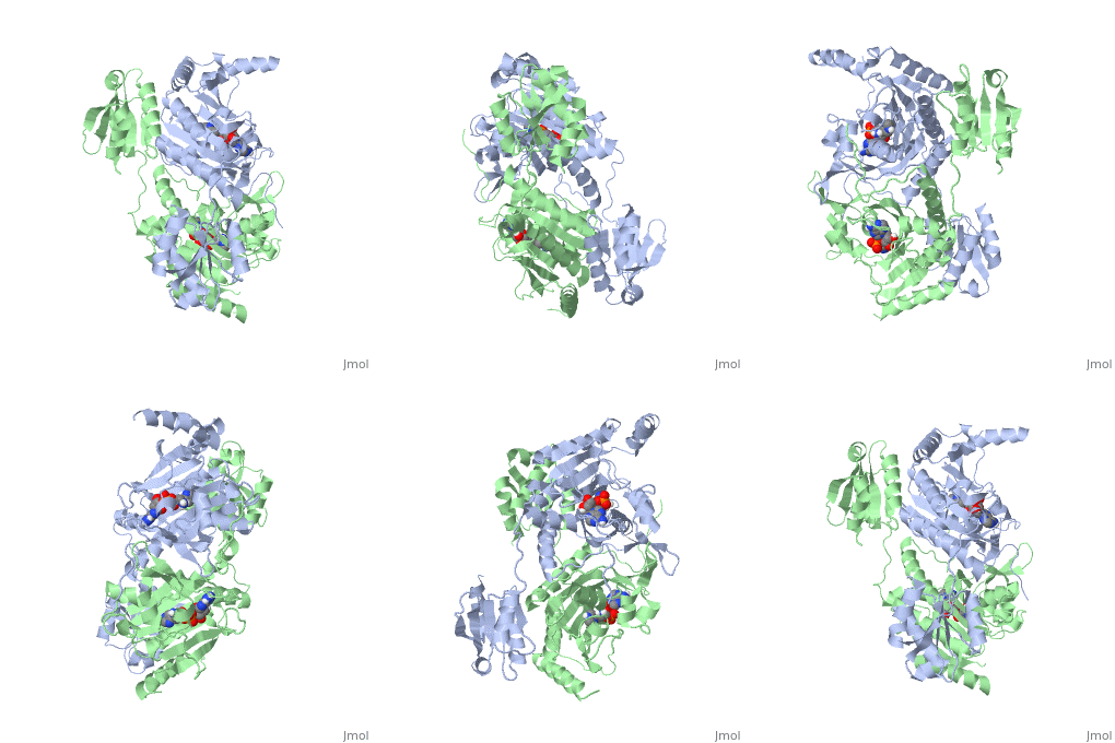
Histidyl tRNA Synthetase (HisRS) is a 94kD that belongs to the class II of aminoacyl-tRNA synthetases (aaRS). Aminoacyl-tRNA synthetases Aminoacyl-tRNA synthetases have been partitioned into two classes, containing 10 members, on the basis of sequence comparisons[1]. Class I and Class II differ mainly with respect to the topology of the catalytic fold and site of esterification on cognate tRNA[1]. Class II enzymes have a composed of anti-parallel β-sheets and α-helices (residues 1-325). Additionally, class II enzymes can be further divided into three subgroups: class IIa, distinguished by an N-terminal catalytic domain and C-terminal accessory domain (later shown to be anticodon binding domain); class IIb, whose anticodon binding domain is located on the N-terminal side of the fold; and class IIc, encompassing the tetrameric PheRS and GlyRS class II synthetases.[2]
| |||||||||||
Mechanism
Evolutionary Conservation
Structural Homology
3D Structures of Histidyl-tRNA Synthetase
Bacteria
Eukaryota
Archara
References
- ↑ 1.0 1.1 Eriani G, Delarue M, Poch O, Gangloff J, Moras D. Partition of tRNA synthetases into two classes based on mutually exclusive sets of sequence motifs. Nature. 1990 Sep 13;347(6289):203-6. PMID:2203971 doi:http://dx.doi.org/10.1038/347203a0
- ↑ Cusack S, Hartlein M, Leberman R. Sequence, structural and evolutionary relationships between class 2 aminoacyl-tRNA synthetases. Nucleic Acids Res. 1991 Jul 11;19(13):3489-98. PMID:1852601
- ↑ 3.0 3.1 3.2 Francklyn, C., and Arnez, J.G. (2004) in Aminoacyl-tRNA Synthetases (Ibba, M.,Francklyn, C.,Cusack, S.. Eds.) Landes Publishing, Austin, TX
- ↑ Arnez JG, Augustine JG, Moras D, Francklyn CS. The first step of aminoacylation at the atomic level in histidyl-tRNA synthetase. Proc Natl Acad Sci U S A. 1997 Jul 8;94(14):7144-9. PMID:9207058
- ↑ Arnez JG, Moras D. Structural and functional considerations of the aminoacylation reaction. Trends Biochem Sci. 1997 Jun;22(6):211-6. PMID:9204708

