Aquaporin
From Proteopedia
| Line 1: | Line 1: | ||
{{STRUCTURE_3gd8| PDB=3gd8 | SIZE=400| SCENE=Aquaporin/Cv/2 |right|CAPTION=Human Aquaporin 4 complex with glycerol and β-octylglucoside, [[3gd8]] }} | {{STRUCTURE_3gd8| PDB=3gd8 | SIZE=400| SCENE=Aquaporin/Cv/2 |right|CAPTION=Human Aquaporin 4 complex with glycerol and β-octylglucoside, [[3gd8]] }} | ||
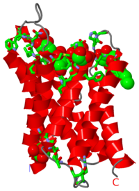
[[Image:3gd8.png|left|200px|thumb|Crystal Structure of Human Aquaporin 4, [[3gd8]]]]'''Aquaporins''' are channel producing proteins which regulate the flow of water across the cell membrane. They are made of α-helix bundles. '''Aquaglyceroporin''' (GLpf) conducts water and polyalcohols. The images at the left and at the right correspond to one representative aquaporin structure, ''i.e.'' the crystal structure of human [[Aquaporin 4]] ([[3gd8]]). The image shows the protein, 6 molecules of glycerol and one of beta-octylglucoside. | [[Image:3gd8.png|left|200px|thumb|Crystal Structure of Human Aquaporin 4, [[3gd8]]]]'''Aquaporins''' are channel producing proteins which regulate the flow of water across the cell membrane. They are made of α-helix bundles. '''Aquaglyceroporin''' (GLpf) conducts water and polyalcohols. The images at the left and at the right correspond to one representative aquaporin structure, ''i.e.'' the crystal structure of human [[Aquaporin 4]] ([[3gd8]]). The image shows the protein, 6 molecules of glycerol and one of beta-octylglucoside. | ||
| + | <br/> | ||
{{TOC limit|limit=2}} | {{TOC limit|limit=2}} | ||
| Line 47: | Line 48: | ||
== 3D Structures of Aquaglyceroporin == | == 3D Structures of Aquaglyceroporin == | ||
| - | |||
[[1lda]], [[1ldi]] – EcGLpf<br /> | [[1lda]], [[1ldi]] – EcGLpf<br /> | ||
[[1ldf]] – EcGLpf (mutant)<br /> | [[1ldf]] – EcGLpf (mutant)<br /> | ||
[[3c02]] – GLpf – ''Plasmodium falciparum''<br /> | [[3c02]] – GLpf – ''Plasmodium falciparum''<br /> | ||
| - | |||
| - | == References == | ||
| - | 1. Crane, J.M., Tajima, M., and Verkman, A.S. Live-cell imaging of aquaporin-4 diffusion and interactions in orthogonal arrays of particles. <i>Neuroscience</i> (2010) vol. 168 (4) pp. 892-902<br/> | ||
| - | 2. Hiroaki, Y., Tani, K., Kamegawa, A., Gyobu, N., Nishikawa, K., Suzuki, H., Walz, T., Sasaki, S., Mitsuoka, Kimura, K., Mizoguchi, A., and Fujiyoshi, Y. Implications of the aquaporin-4 structure on array formation and cell adhesion. <i>J Mol Biol</i> (2006) vol. 355 (4) pp. 628-39<br/> | ||
| - | 3. Nicchia, G.P., Rossi, A., Mola, M.G., Pisani, F., Stigliano, C., Basco, D., Mastrototaro, M., Svelto, M., and A. Frigeri. Higher order structure of aquaporin-4. <i>Neuroscience</i> (2010) vol. 168 (4) pp. 903-14<br/> | ||
| - | 4. Pittock SJ, and Lennon V.A. Aquaporin-4 autoantibodies in a paraneoplastic context. <i>Arch Neurol</i> (2008) 65:629–632.<br/> | ||
| - | 5. Hinson, S.R., McKeon, A., and Lennon V.A. Neurological autoimmunity targeting aquaporin-4. <i>Neuroscience</i> (2010) vol. 168 (4) pp. 1009-18.<br/> | ||
| - | 6. Saadoun S., Papadopoulos M.C., Hara-Chikuma M, Verkman A.S. Impairment of angiogenesis and cell migration by targeted aquaporin-1 gene disruption. <i>Nature</i> (2005) 434:786–792.<br/> | ||
| - | 7. Verkman A.S., Hara-Chikuma M., Papadopoulos M.C. Aquaporins—new players in cancer biology. <i>J Mol Med</i> (2008) 86:523–529. | ||
Revision as of 01:46, 27 June 2012
| |||||||||
| Human Aquaporin 4 complex with glycerol and β-octylglucoside, 3gd8 | |||||||||
|---|---|---|---|---|---|---|---|---|---|
| Ligands: | , | ||||||||
| Gene: | AQP4 (Homo sapiens) | ||||||||
| |||||||||
| |||||||||
| Resources: | FirstGlance, OCA, RCSB, PDBsum | ||||||||
| Coordinates: | save as pdb, mmCIF, xml | ||||||||

Contents |
3D Structures of Aquaporin
Aquaporin
3llq – Aqp – Agrobacterium tumefaciens
3cll, 3cn5, 3cn6 – sAqp SoPIP2 (mutant) – spinach
1z98, 2b5f - sAqp SoPIP2
Aquaporin 0
2c32, 1ymg, 2b6p – cAqp0 – cow
2b6o – Aqp0 - electron crystallography – sheep
1sor, 3m9i – Aqp0 – Ovis aries
Aquaporin 1
2w1p, 2w2e – Aqp1 – Pischia pastoris
1fqy – hAqp1 – electron crystallography - human
1h6i, 1ih5 – hAqp1
1j4n – cAqp1
Aquaporin 4
2zz9 – rAqp4 (mutant) – rat
2d57, 3iyz – rAqp4 – electron crystallography
3gd8 – hAqp4 – human
Aquaporin 5
3d9s – hAqp5
Aquaporin M
2evu, 2f2b – AqpM – Methanothermobacter marburgensis
Aquaporin Z
2o9d, 2o9f, 3nk5, 3nka, 3nkc – EcAqpZ (mutant) – Escherichia coli
2o9e, 2o9g - EcAqpZ (mutant)+Hg
2abm, 1rc2 - EcAqpZ
3D Structures of Aquaglyceroporin
1lda, 1ldi – EcGLpf
1ldf – EcGLpf (mutant)
3c02 – GLpf – Plasmodium falciparum
Proteopedia Page Contributors and Editors (what is this?)
Michal Harel, Alexander Berchansky, Mark Leiserson, David Canner, Jaime Prilusky, Eran Hodis

