Journal:JBSD:18
From Proteopedia
(Difference between revisions)

(New page: .) |
|||
| (12 intermediate revisions not shown.) | |||
| Line 1: | Line 1: | ||
| - | . | + | <StructureSection load='' size='450' side='right' scene='Journal:JBSD:18/Cv/3' caption=''> |
| + | === Molecular modeling study for conformational changes of Sirtuin 2 due to substrate and inhibitor binding === | ||
| + | <big>Sugunadevi Sakkiah, Meganathan Chandrasekaran, Yuno Lee, Songmi Kim, Keun Woo Lee </big> <ref>DOI 10.1080/07391102.2012.680026</ref> | ||
| + | <hr/> | ||
| + | <b>Molecular Tour</b><br> | ||
| + | <scene name='Journal:JBSD:18/Cv/4'>Sirtuin</scene>, belongs to the member of NAD+-dependent deacetylase family. Structural detail of sirtuin 2 (SIRT2) complexes will be most utilitarian to discover the drug which might have a beneficial effects on a variety of human diseases like cancer, diabetes etc. Unfortunately till now there is a lack of SIRT2-ligand complex structure details, hence molecular docking was carried out to dock the substrate (NAD and acetylated lysine) and inhibitor (sirtinol) in the NAD binding pocket. The suitable binding orientation of <scene name='Journal:JBSD:18/Cv/8'>substrate</scene> (<span style="color:yellow;background-color:black;font-weight:bold;">protein is colored in yellow</span>, <font color='magenta'><b>NAD is in magenta</b></font>) and <scene name='Journal:JBSD:18/Cv/9'>inhibitor</scene> (<span style="color:lime;background-color:black;font-weight:bold;">protein is colored in green</span>, <span style="color:salmon;background-color:black;font-weight:bold;">inhibitor is in salmon</span>), in SIRT2 active site were subjected to 5 ns molecular dynamics simulations to adjust the docked ligand structure into the active site and to identify its <scene name='Journal:JBSD:18/Cv/10'>conformational changes</scene> and dynamic behavior in active site. The result of our study affords an idea about the 3D structural details of SIRT2 in presence of substrate and inhibitor and their orientation in active site, the <scene name='Journal:JBSD:18/Cv/11'>importance of F96 was unveiled</scene>. In addition, the simulation revealed that the displacement of F96 upon substrate and inhibitor binding induced an <scene name='Journal:JBSD:18/Cv/14'>extended conformation of loop3</scene> and <scene name='Journal:JBSD:18/Cv/16'>changes its interactions with the rest of SIRT2</scene>. We believed that our SIRT2 modeling studies could be much helpful to gain the structural insight of SIRT2 and also this will be useful to design the receptor-based inhibitors. | ||
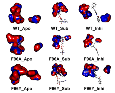
| + | [[Image:JBSD18.png|left|400px|thumb|Electrostatic potential map indicates the assembly and disassembly of C pocket in the presence of NAD+ and inhibitor. Apo-form (left column), SIRT2–substrate (central column), and SIRT2–inhibitor (right column).]] | ||
| + | |||
| + | |||
| + | </StructureSection> | ||
| + | |||
| + | <references/> | ||
| + | __NOEDITSECTION__ | ||
Current revision
| |||||||||||
- ↑ Sakkiah S, Chandrasekaran M, Lee Y, Kim S, Lee KW. Molecular modeling study for conformational changes of Sirtuin 2 due to substrate and inhibitor binding. J Biomol Struct Dyn. 2012 Jul;30(3):235-54. Epub 2012 Jun 12. PMID:22694102 doi:10.1080/07391102.2012.680026
This page complements a publication in scientific journals and is one of the Proteopedia's Interactive 3D Complement pages. For aditional details please see I3DC.

