This old version of Proteopedia is provided for student assignments while the new version is undergoing repairs. Content and edits done in this old version of Proteopedia after March 1, 2026 will eventually be lost when it is retired in about June of 2026.
Apply for new accounts at the new Proteopedia. Your logins will work in both the old and new versions.
1rss
From Proteopedia
(New page: 200px<br /><applet load="1rss" size="450" color="white" frame="true" align="right" spinBox="true" caption="1rss, resolution 1.9Å" /> '''RIBOSOMAL PROTEIN S7 ...) |
|||
| Line 1: | Line 1: | ||
| - | [[Image:1rss.gif|left|200px]]<br /><applet load="1rss" size=" | + | [[Image:1rss.gif|left|200px]]<br /><applet load="1rss" size="350" color="white" frame="true" align="right" spinBox="true" |
caption="1rss, resolution 1.9Å" /> | caption="1rss, resolution 1.9Å" /> | ||
'''RIBOSOMAL PROTEIN S7 FROM THERMUS THERMOPHILUS'''<br /> | '''RIBOSOMAL PROTEIN S7 FROM THERMUS THERMOPHILUS'''<br /> | ||
==Overview== | ==Overview== | ||
| - | BACKGROUND: Ribosomal protein S7, a crucial RNA-binding component of the | + | BACKGROUND: Ribosomal protein S7, a crucial RNA-binding component of the ribosome, is one of two proteins that initiates assembly of the 30S ribosomal subunit. It is required for proper folding of a large 3' domain of 16S ribosomal RNA. S7 regulates its own synthesis by binding to its own mRNA. This ability of S7 to bind both messenger and ribosomal RNAs makes determination of its mode of RNA recognition particularly interesting. RESULTS: The crystal structure of S7 from Thermus thermophilus was determined by a two-wavelength anomalous diffraction experiment using the LIII edge of mercury. The S7 structure consists of a bundle of six helices and an extended beta hairpin between helices 3 and 4, with two or more RNA-binding sites on its surface. The hairpin, along with portions of helices 1, 4 and 6, forms a large, positively charged, concave surface that has the appropriate curvature and dimensions to bind double-stranded RNA. A second putative RNA-binding site comprises parts of loop 2 and the helix 4-loop 5 turn. CONCLUSIONS: Structural similarity between S7 and the IHF/HU family of proteins strongly suggests that the beta hairpin of S7 binds to a groove of double-stranded RNA. The beta hairpin of S7 is also similar to those from other nucleic acid binding proteins, such as ribosomal protein L14 and BIV Tat, suggesting that it belongs to an extended family of such motifs, all of which bind to a groove of double-stranded nucleic acid. The residues in S7 loop 2 that belong to the second putative RNA-binding site may have a role analogous to the N-terminal residues of IHF/HU which grip an unbent portion of double helix. |
==About this Structure== | ==About this Structure== | ||
| - | 1RSS is a [http://en.wikipedia.org/wiki/Single_protein Single protein] structure of sequence from [http://en.wikipedia.org/wiki/Thermus_thermophilus Thermus thermophilus]. Full crystallographic information is available from [http:// | + | 1RSS is a [http://en.wikipedia.org/wiki/Single_protein Single protein] structure of sequence from [http://en.wikipedia.org/wiki/Thermus_thermophilus Thermus thermophilus]. Full crystallographic information is available from [http://oca.weizmann.ac.il/oca-bin/ocashort?id=1RSS OCA]. |
==Reference== | ==Reference== | ||
| Line 20: | Line 20: | ||
[[Category: translation]] | [[Category: translation]] | ||
| - | ''Page seeded by [http:// | + | ''Page seeded by [http://oca.weizmann.ac.il/oca OCA ] on Thu Feb 21 14:54:06 2008'' |
Revision as of 12:54, 21 February 2008
|
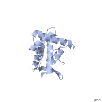
RIBOSOMAL PROTEIN S7 FROM THERMUS THERMOPHILUS
Overview
BACKGROUND: Ribosomal protein S7, a crucial RNA-binding component of the ribosome, is one of two proteins that initiates assembly of the 30S ribosomal subunit. It is required for proper folding of a large 3' domain of 16S ribosomal RNA. S7 regulates its own synthesis by binding to its own mRNA. This ability of S7 to bind both messenger and ribosomal RNAs makes determination of its mode of RNA recognition particularly interesting. RESULTS: The crystal structure of S7 from Thermus thermophilus was determined by a two-wavelength anomalous diffraction experiment using the LIII edge of mercury. The S7 structure consists of a bundle of six helices and an extended beta hairpin between helices 3 and 4, with two or more RNA-binding sites on its surface. The hairpin, along with portions of helices 1, 4 and 6, forms a large, positively charged, concave surface that has the appropriate curvature and dimensions to bind double-stranded RNA. A second putative RNA-binding site comprises parts of loop 2 and the helix 4-loop 5 turn. CONCLUSIONS: Structural similarity between S7 and the IHF/HU family of proteins strongly suggests that the beta hairpin of S7 binds to a groove of double-stranded RNA. The beta hairpin of S7 is also similar to those from other nucleic acid binding proteins, such as ribosomal protein L14 and BIV Tat, suggesting that it belongs to an extended family of such motifs, all of which bind to a groove of double-stranded nucleic acid. The residues in S7 loop 2 that belong to the second putative RNA-binding site may have a role analogous to the N-terminal residues of IHF/HU which grip an unbent portion of double helix.
About this Structure
1RSS is a Single protein structure of sequence from Thermus thermophilus. Full crystallographic information is available from OCA.
Reference
The structure of ribosomal protein S7 at 1.9 A resolution reveals a beta-hairpin motif that binds double-stranded nucleic acids., Wimberly BT, White SW, Ramakrishnan V, Structure. 1997 Sep 15;5(9):1187-98. PMID:9331418
Page seeded by OCA on Thu Feb 21 14:54:06 2008
