This old version of Proteopedia is provided for student assignments while the new version is undergoing repairs. Content and edits done in this old version of Proteopedia after March 1, 2026 will eventually be lost when it is retired in about June of 2026.
Apply for new accounts at the new Proteopedia. Your logins will work in both the old and new versions.
Single stranded binding protein
From Proteopedia
| Line 16: | Line 16: | ||
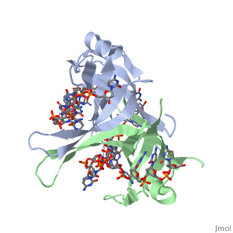
Single-stranded DNA can interact with SSB through hydrogen bonds, stacking, or electronegative interactions. Though SSB proteins are found in a variety of different organisms, most interactions between SSB and ssDNA happen through the common structural motif of an oligosaccharide/oligonucleotide binding site, referred to as the OB fold <ref>Shamoo, Yousif. “Single Stranded DNA binding proteins.” ‘’Encyclopedia of Life Sciences.’’ MacMillan Publishers Ltd, Nature Publishing Group; 2002</ref>. The OB fold allows SSB to bind preferentially to ssDNA. Each subunit of a SSB has an OB fold (the SSB of E. coli thus has four OB folds, one per each of its four identical subunits). This fold consists of a 5-stranded β barrel that ends in an α-helix. | Single-stranded DNA can interact with SSB through hydrogen bonds, stacking, or electronegative interactions. Though SSB proteins are found in a variety of different organisms, most interactions between SSB and ssDNA happen through the common structural motif of an oligosaccharide/oligonucleotide binding site, referred to as the OB fold <ref>Shamoo, Yousif. “Single Stranded DNA binding proteins.” ‘’Encyclopedia of Life Sciences.’’ MacMillan Publishers Ltd, Nature Publishing Group; 2002</ref>. The OB fold allows SSB to bind preferentially to ssDNA. Each subunit of a SSB has an OB fold (the SSB of E. coli thus has four OB folds, one per each of its four identical subunits). This fold consists of a 5-stranded β barrel that ends in an α-helix. | ||
| - | Several specific amino acid residues play essential roles in the binding of ssDNA to SSB. <scene name='56/566528/Phe_60/ | + | Several specific amino acid residues play essential roles in the binding of ssDNA to SSB. <scene name='56/566528/Phe_60/2'>Phe60</scene> is a key residue involved in binding the ssDNA to the protein, as it has been shown to be the site for cross-linking. Tryptophan and Lysine residues are important in binding as well, as evidenced by modification treatments of lysine and tryptophan residues resulting a complete loss of binding activity for the protein. The two tryptophan residues involved in ssDNA binding are Trp40 and Trp54, which were determined by mutagenesis <ref>PMID:2087220</ref>. |
One more key residue in the binding site, His55, was determined by site-specific mutagenesis, as when His55 is substituted with Leu it decreases the overall binding affinity for ssDNA. All of these residues are found in a hydrophobic region, which is suitable for nucleotide base interactions. Treatments that modified arginine, cysteine, or tyrosine residues had no effect on binding of SSB to DNA, suggesting that these amino acids are not involved in significant interactions of the protein with the ssDNA. | One more key residue in the binding site, His55, was determined by site-specific mutagenesis, as when His55 is substituted with Leu it decreases the overall binding affinity for ssDNA. All of these residues are found in a hydrophobic region, which is suitable for nucleotide base interactions. Treatments that modified arginine, cysteine, or tyrosine residues had no effect on binding of SSB to DNA, suggesting that these amino acids are not involved in significant interactions of the protein with the ssDNA. | ||
Revision as of 01:03, 3 November 2013
Sandbox Single Stranded DNA-Binding Protein (SSB)
Contents |
Overview
Single-stranded DNA-binding protein, or SSB binds to single-stranded regions of DNA. This binding serves a variety of functions - it prevents the strands from hardening too early during replication, it protects the single-stranded DNA from being broken down by nucleases during repair, and it removes the secondary structure of the strands so that other enzymes are able to access them and act effectively upon the strands[1].
Single-stranded DNA (ssDNA) is utilized primarily during the course of major aspects of DNA metabolism such as replication, recombination and repair [2]. In addition to stabilizing ssDNA, SSB proteins also bind to and control the function of many other proteins that are involved in all of three of these major DNA metabolic processes. During DNA replication, SSB molecules bind to the newly separated individual DNA strands, keeping the strands separated by holding them in place so that each strand can serve as a template for new DNA synthesis[3].
Structure of E. coli SSB
| |||||||||||
Binding Interactions in the Active Site
Single-stranded DNA can interact with SSB through hydrogen bonds, stacking, or electronegative interactions. Though SSB proteins are found in a variety of different organisms, most interactions between SSB and ssDNA happen through the common structural motif of an oligosaccharide/oligonucleotide binding site, referred to as the OB fold [10]. The OB fold allows SSB to bind preferentially to ssDNA. Each subunit of a SSB has an OB fold (the SSB of E. coli thus has four OB folds, one per each of its four identical subunits). This fold consists of a 5-stranded β barrel that ends in an α-helix.
Several specific amino acid residues play essential roles in the binding of ssDNA to SSB. is a key residue involved in binding the ssDNA to the protein, as it has been shown to be the site for cross-linking. Tryptophan and Lysine residues are important in binding as well, as evidenced by modification treatments of lysine and tryptophan residues resulting a complete loss of binding activity for the protein. The two tryptophan residues involved in ssDNA binding are Trp40 and Trp54, which were determined by mutagenesis [11].
One more key residue in the binding site, His55, was determined by site-specific mutagenesis, as when His55 is substituted with Leu it decreases the overall binding affinity for ssDNA. All of these residues are found in a hydrophobic region, which is suitable for nucleotide base interactions. Treatments that modified arginine, cysteine, or tyrosine residues had no effect on binding of SSB to DNA, suggesting that these amino acids are not involved in significant interactions of the protein with the ssDNA.
Interactions Between E. coli SSB and other Proteins
Most of the molecule loses flexibility after ssDNA binding. However, three phenylalanine residues (Phe147, Phe171, Phe177) in the COOH terminal domain remain flexible, even after DNA binding, suggesting that the COOH terminus has something to do with protein binding [12]. An experiment where Phe177 was changed to Cys resulted in a protein that could not replicate DNA. This replication defect stemming from the lost phenylalanine residue was likely a result of the inability of the altered C-terminal region to bind other proteins necessary for replication [13]. Gly15 is believed to play an important role in binding the RecA protein. Mutations in Gly15 have been shown to have extreme effects on recombinational repair. SSB is also thought to bind with exonuclease I, DNA polymerase II, and a protein n, which is a part of the primosome complex and used to help synthesize RNA primers for the lagging strand [14].
Other SSB Structures
| |||||||||||
See Also
References
- ↑ Meyer RR, Laine PS. The single-stranded DNA-binding protein of Escherichia coli. Microbiol Rev. 1990 Dec;54(4):342-80. PMID:2087220
- ↑ Meyer RR, Laine PS. The single-stranded DNA-binding protein of Escherichia coli. Microbiol Rev. 1990 Dec;54(4):342-80. PMID:2087220
- ↑ Berg JM, Tymoczko JL, Stryer L. Biochemistry. 6th edition. New York: W H Freeman; 2006.
- ↑ Meyer RR, Laine PS. The single-stranded DNA-binding protein of Escherichia coli. Microbiol Rev. 1990 Dec;54(4):342-80. PMID:2087220
- ↑ Kozlov AG, Lohman TM. Stopped-flow studies of the kinetics of single-stranded DNA binding and wrapping around the Escherichia coli SSB tetramer. Biochemistry. 2002 May 14;41(19):6032-44. PMID:11993998
- ↑ Kozlov AG, Lohman TM. Stopped-flow studies of the kinetics of single-stranded DNA binding and wrapping around the Escherichia coli SSB tetramer. Biochemistry. 2002 May 14;41(19):6032-44. PMID:11993998
- ↑ Kozlov AG, Lohman TM. Stopped-flow studies of the kinetics of single-stranded DNA binding and wrapping around the Escherichia coli SSB tetramer. Biochemistry. 2002 May 14;41(19):6032-44. PMID:11993998
- ↑ Meyer RR, Laine PS. The single-stranded DNA-binding protein of Escherichia coli. Microbiol Rev. 1990 Dec;54(4):342-80. PMID:2087220
- ↑ Meyer RR, Laine PS. The single-stranded DNA-binding protein of Escherichia coli. Microbiol Rev. 1990 Dec;54(4):342-80. PMID:2087220
- ↑ Shamoo, Yousif. “Single Stranded DNA binding proteins.” ‘’Encyclopedia of Life Sciences.’’ MacMillan Publishers Ltd, Nature Publishing Group; 2002
- ↑ Meyer RR, Laine PS. The single-stranded DNA-binding protein of Escherichia coli. Microbiol Rev. 1990 Dec;54(4):342-80. PMID:2087220
- ↑ Meyer RR, Laine PS. The single-stranded DNA-binding protein of Escherichia coli. Microbiol Rev. 1990 Dec;54(4):342-80. PMID:2087220
- ↑ Agamova KA, Gladunova ZD, Savinkin IuN. [Cytologic method in the diagnosis of precancerous conditions and early cancer of the stomach]. Lab Delo. 1988;(3):43-5. PMID:2453719
- ↑ Meyer RR, Laine PS. The single-stranded DNA-binding protein of Escherichia coli. Microbiol Rev. 1990 Dec;54(4):342-80. PMID:2087220
Proteopedia Page Contributors and Editors (what is this?)
Refayat Ahsen, Rachel Craig, Michal Harel, Alexander Berchansky
