This old version of Proteopedia is provided for student assignments while the new version is undergoing repairs. Content and edits done in this old version of Proteopedia after March 1, 2026 will eventually be lost when it is retired in about June of 2026.
Apply for new accounts at the new Proteopedia. Your logins will work in both the old and new versions.
Sanbox glut3
From Proteopedia
(Difference between revisions)
| Line 8: | Line 8: | ||
==Structure== | ==Structure== | ||
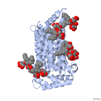
| - | <scene name='pdbligand=37X:OCTYL+GLUCOSE+NEOPENTYL+GLYCOL'>37X</scene>, <scene name='pdbligand=Y01:CHOLESTEROL+HEMISUCCINATE'>Y01</scene> | + | GLUT3(5c65) is a transport protein consisting of 481 amino acids and weighing 52,520 Daltons in its asymmetrical unit. This protein is an alpha-helical protein consisting of two chains, two different ligands and water. The structure was determined by X-Ray diffraction and was measured at a resolution of 2.65 Angstroms. GLUT3 consists of 12 transmembrane segments (TMs) folded “into the N-terminal and C-terminal domains, each comprising ‘3+3’ inverted repeats” These TMs consist of four 3 repeated sections (figure 1 part d). The protein consists of two different ligands, Y01(green) and 37X(green). Octyl Glucose Neopentyl Glycol (<scene name='pdbligand=37X:OCTYL+GLUCOSE+NEOPENTYL+GLYCOL'>37X</scene>) has a chemical formula of C27H52O12 and a molecular weight of 569 Da. There are six 37X (501-506a) bound to chain A of 5c65. These ligands are kept in place by hydrogen bonds to arginine, proline, and serine and by van der Waals forces. Chain B has three 37X ligands attached to it (501-503b). These are attached through hydrogen bonds by arginine, proline, and serine as well as by van der Waals forces. Cholesterol hemisuccinate (<scene name='pdbligand=Y01:CHOLESTEROL+HEMISUCCINATE'>Y01</scene>) has a chemical formula of C31H50O4 and has a molecular weight of 487 Da. One Y01 is attached to chain a and another Y01 is attached to chain b. GLUT3 was also identified and analyzed in a complex with alpha & beta d-glucose. This model was reported with a resolution of 1.5 Å and was in an open-occluded state. The alpha and beta d glucose were coordinated by amino acids N315, E378, Q159, W368, Q280, Q281, N286. These are located on TM8 and TM10a and TM10b. GLUT3 structure was also determined when bound to maltose in an outward-open and an outward-occluded conformation. This was measure to a resolution of 2.6 Å and 2.4 Å respectively. (include Figure 3 part a). To get a better view of the structure of the protein use FirstGlance (link). |
| + | |||
== Disease in Humans == | == Disease in Humans == | ||
Alzheimer’s disease, shows levels of impaired glucose uptake and metabolism, which leads to the downgrade of many other factors in the brain. GLUT3 is responsible for transporting glucose from extracellular space to neuronal tissue (i.e. dendrites and axons). Decreased levels of GLUT3 in Alzheimer brain shows a positive correlation to decreased levels of N-acetylglucosamine. The impaired presence of GLUT3 leads to hyperphosphorylation of the Tau protein, which normally stabilizes neuronal microtubules. Lastly there is a reduction in the transcription for factor hypoxia-inducible factor 1, which plays a role in glucose metabolism in the brain. The comparison between a normal healthy brain and an Alzheimer brain relieved that there was a 25-30% decrease in GLUT3 levels in the Alzheimer brain. | Alzheimer’s disease, shows levels of impaired glucose uptake and metabolism, which leads to the downgrade of many other factors in the brain. GLUT3 is responsible for transporting glucose from extracellular space to neuronal tissue (i.e. dendrites and axons). Decreased levels of GLUT3 in Alzheimer brain shows a positive correlation to decreased levels of N-acetylglucosamine. The impaired presence of GLUT3 leads to hyperphosphorylation of the Tau protein, which normally stabilizes neuronal microtubules. Lastly there is a reduction in the transcription for factor hypoxia-inducible factor 1, which plays a role in glucose metabolism in the brain. The comparison between a normal healthy brain and an Alzheimer brain relieved that there was a 25-30% decrease in GLUT3 levels in the Alzheimer brain. | ||
Revision as of 03:53, 17 November 2015
Facilitated Glucose Transporter 3, Solute Carrier Family 2 (GLUT3/ SLC2A3) in Homo Sapiens
| |||||||||||
References
- ↑ Hanson, R. M., Prilusky, J., Renjian, Z., Nakane, T. and Sussman, J. L. (2013), JSmol and the Next-Generation Web-Based Representation of 3D Molecular Structure as Applied to Proteopedia. Isr. J. Chem., 53:207-216. doi:http://dx.doi.org/10.1002/ijch.201300024
- ↑ Herraez A. Biomolecules in the computer: Jmol to the rescue. Biochem Mol Biol Educ. 2006 Jul;34(4):255-61. doi: 10.1002/bmb.2006.494034042644. PMID:21638687 doi:10.1002/bmb.2006.494034042644
- ↑ 3.0 3.1 Long, W., & Cheeseman, C. I. (2015). Structure of, and functional insight into the GLUT family of membrane transporters. Cell Health and Cytoskeleton, 7, 167-183. doi:10.2147/CHC.S60484
