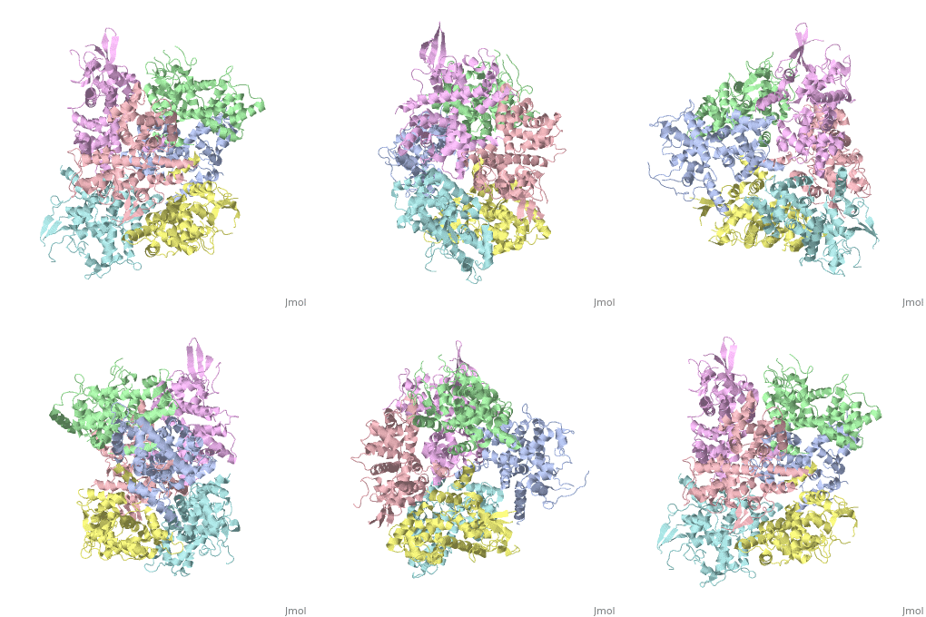
Calcium uptake protein 1
From Proteopedia
(Difference between revisions)
| Line 19: | Line 19: | ||
== Structural highlights == | == Structural highlights == | ||
Ca 2+ binds to EF domain on MICU 1 and 2 | Ca 2+ binds to EF domain on MICU 1 and 2 | ||
| - | <scene name='72/723172/Protein_dimer_when_ca_is_bound/ | + | <scene name='72/723172/Protein_dimer_when_ca_is_bound/3'>Here is the binding site of calcium in the dimer</scene> |
| + | |||
This is a sample scene created with SAT to <scene name="/12/3456/Sample/1">color</scene> by Group, and another to make <scene name="/12/3456/Sample/2">a transparent representation</scene> of the protein. You can make your own scenes on SAT starting from scratch or loading and editing one of these sample scenes. | This is a sample scene created with SAT to <scene name="/12/3456/Sample/1">color</scene> by Group, and another to make <scene name="/12/3456/Sample/2">a transparent representation</scene> of the protein. You can make your own scenes on SAT starting from scratch or loading and editing one of these sample scenes. | ||
Revision as of 13:37, 28 January 2016
Your Heading Here (maybe something like 'Structure')
| |||||||||||

