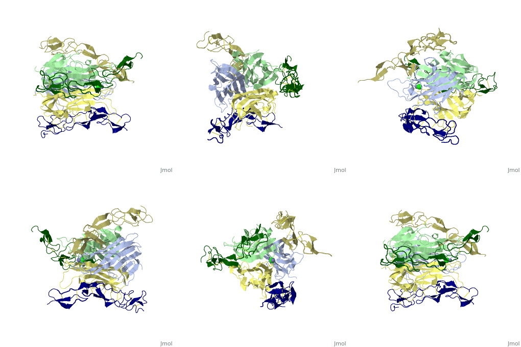
1d0g
From Proteopedia
| Line 4: | Line 4: | ||
|PDB= 1d0g |SIZE=350|CAPTION= <scene name='initialview01'>1d0g</scene>, resolution 2.4Å | |PDB= 1d0g |SIZE=350|CAPTION= <scene name='initialview01'>1d0g</scene>, resolution 2.4Å | ||
|SITE= | |SITE= | ||
| - | |LIGAND= <scene name='pdbligand= | + | |LIGAND= <scene name='pdbligand=CL:CHLORIDE+ION'>CL</scene>, <scene name='pdbligand=ZN:ZINC+ION'>ZN</scene> |
|ACTIVITY= | |ACTIVITY= | ||
|GENE= | |GENE= | ||
| + | |DOMAIN= | ||
| + | |RELATEDENTRY= | ||
| + | |RESOURCES=<span class='plainlinks'>[http://oca.weizmann.ac.il/oca-docs/fgij/fg.htm?mol=1d0g FirstGlance], [http://oca.weizmann.ac.il/oca-bin/ocaids?id=1d0g OCA], [http://www.ebi.ac.uk/pdbsum/1d0g PDBsum], [http://www.rcsb.org/pdb/explore.do?structureId=1d0g RCSB]</span> | ||
}} | }} | ||
| Line 14: | Line 17: | ||
==Overview== | ==Overview== | ||
Formation of a complex between Apo2L (also called TRAIL) and its signaling receptors, DR4 and DR5, triggers apoptosis by inducing the oligomerization of intracellular death domains. We report the crystal structure of the complex between Apo2L and the ectodomain of DR5. The structure shows three elongated receptors snuggled into long crevices between pairs of monomers of the homotrimeric ligand. The interface is divided into two distinct patches, one near the bottom of the complex close to the receptor cell surface and one near the top. Both patches contain residues that are critical for high-affinity binding. A comparison to the structure of the lymphotoxin-receptor complex suggests general principles of binding and specificity for ligand recognition in the TNF receptor superfamily. | Formation of a complex between Apo2L (also called TRAIL) and its signaling receptors, DR4 and DR5, triggers apoptosis by inducing the oligomerization of intracellular death domains. We report the crystal structure of the complex between Apo2L and the ectodomain of DR5. The structure shows three elongated receptors snuggled into long crevices between pairs of monomers of the homotrimeric ligand. The interface is divided into two distinct patches, one near the bottom of the complex close to the receptor cell surface and one near the top. Both patches contain residues that are critical for high-affinity binding. A comparison to the structure of the lymphotoxin-receptor complex suggests general principles of binding and specificity for ligand recognition in the TNF receptor superfamily. | ||
| - | |||
| - | ==Disease== | ||
| - | Known diseases associated with this structure: Squamous cell carcinoma, head and neck OMIM:[[http://www.ncbi.nlm.nih.gov/entrez/dispomim.cgi?id=603612 603612]] | ||
==About this Structure== | ==About this Structure== | ||
| Line 32: | Line 32: | ||
[[Category: Kelley, R F.]] | [[Category: Kelley, R F.]] | ||
[[Category: Vos, A M.de.]] | [[Category: Vos, A M.de.]] | ||
| - | [[Category: CL]] | ||
| - | [[Category: ZN]] | ||
[[Category: apoptosis]] | [[Category: apoptosis]] | ||
[[Category: binding and specificity]] | [[Category: binding and specificity]] | ||
| Line 39: | Line 37: | ||
[[Category: tnf receptor family]] | [[Category: tnf receptor family]] | ||
| - | ''Page seeded by [http://oca.weizmann.ac.il/oca OCA ] on | + | ''Page seeded by [http://oca.weizmann.ac.il/oca OCA ] on Sun Mar 30 19:32:02 2008'' |
Revision as of 16:32, 30 March 2008
| |||||||
| , resolution 2.4Å | |||||||
|---|---|---|---|---|---|---|---|
| Ligands: | , | ||||||
| Resources: | FirstGlance, OCA, PDBsum, RCSB | ||||||
| Coordinates: | save as pdb, mmCIF, xml | ||||||
CRYSTAL STRUCTURE OF DEATH RECEPTOR 5 (DR5) BOUND TO APO2L/TRAIL
Overview
Formation of a complex between Apo2L (also called TRAIL) and its signaling receptors, DR4 and DR5, triggers apoptosis by inducing the oligomerization of intracellular death domains. We report the crystal structure of the complex between Apo2L and the ectodomain of DR5. The structure shows three elongated receptors snuggled into long crevices between pairs of monomers of the homotrimeric ligand. The interface is divided into two distinct patches, one near the bottom of the complex close to the receptor cell surface and one near the top. Both patches contain residues that are critical for high-affinity binding. A comparison to the structure of the lymphotoxin-receptor complex suggests general principles of binding and specificity for ligand recognition in the TNF receptor superfamily.
About this Structure
1D0G is a Protein complex structure of sequences from Homo sapiens. Full crystallographic information is available from OCA.
Reference
Triggering cell death: the crystal structure of Apo2L/TRAIL in a complex with death receptor 5., Hymowitz SG, Christinger HW, Fuh G, Ultsch M, O'Connell M, Kelley RF, Ashkenazi A, de Vos AM, Mol Cell. 1999 Oct;4(4):563-71. PMID:10549288
Page seeded by OCA on Sun Mar 30 19:32:02 2008

