Z-DNA model tour
From Proteopedia
(Difference between revisions)
| Line 30: | Line 30: | ||
Now change the display to make it show the <scene name='72/725870/Space_filling_bbone/1'>sugar-phosphate backbone as pseudo-bonds</scene> connecting the phosphate atoms. Now the bases are easier to see. Now the bases are easier to see. Notice how they are stacked upon each other and are nearly perpendicular to the axis of the double helix. But notice that the base pairs do not stack upon each other equivalently. The backbone also is not a continuous curve, it "zig-zags" back and forth (hence "Z"-DNA). | Now change the display to make it show the <scene name='72/725870/Space_filling_bbone/1'>sugar-phosphate backbone as pseudo-bonds</scene> connecting the phosphate atoms. Now the bases are easier to see. Now the bases are easier to see. Notice how they are stacked upon each other and are nearly perpendicular to the axis of the double helix. But notice that the base pairs do not stack upon each other equivalently. The backbone also is not a continuous curve, it "zig-zags" back and forth (hence "Z"-DNA). | ||
| - | + | <scene name='72/725870/Zoom_pair/1'>In this view</scene>, the molecule is shown in stick representation, with the backbone in yellow and sets of base pairs in red and blue. Notice how the blue bases stack well on the adjacent blue ones, but not on adjacent red ones, and vice versa. So it is the dinucleotide unit, rather than mononucleotide that is the repeating unit of the structure. This explains the need for alternating purines and pyrimidines to form Z-DNA. | |
| - | + | ||
| - | + | You can see <scene name='72/725870/Zoom_pairs_only/1'>the same view without the backbone</scene> here.Going 5' to 3', there is good stacking within the GpC dinucleotide, but not between them (CpG). | |
| - | + | A also illustrates the stacking arrangement. | |
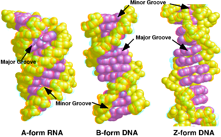
| - | You can compare it with the DNA forms by looking at this [http://proteopedia.org/wiki/images/d/d3/JnABZ3d.gif 3D red-blue | + | . You can also see this |
| + | You can compare it with the DNA forms by looking at this [http://proteopedia.org/wiki/images/d/d3/JnABZ3d.gif 3D red-blue stereo picture of A, B, and Z DNA] | ||
</StructureSection> | </StructureSection> | ||
== References == | == References == | ||
Revision as of 20:46, 21 February 2016
Z-form DNA model
| |||||||||||
References
R. E. Dickerson, H. R. Drew, B. N. Conner, R. M. Wing, A. V. Fratini & M. L. Kopka (1982) The anatomy of A-, B-, and Z-DNA. Science 216: 475-485 [1] JSmol in Proteopedia [2] or to the article describing Jmol [3] to the rescue.
