We apologize for Proteopedia being slow to respond. For the past two years, a new implementation of Proteopedia has been being built. Soon, it will replace this 18-year old system. All existing content will be moved to the new system at a date that will be announced here.
User:Daniel Schemenauer/Sandbox 1
From Proteopedia
(Difference between revisions)
| Line 1: | Line 1: | ||
| - | <StructureSection load='4oo9' size='340' side='right' caption='metabotropic Glutamate | + | <StructureSection load='4oo9' size='340' side='right' caption='metabotropic Glutamate Receptor 5 PDB:[http://www.rcsb.org/pdb/explore/explore.do?structureId=4oo9 4oo9]' scene='72/726409/Overview/5'> |
= metabotropic Glutamate Receptor 5 = | = metabotropic Glutamate Receptor 5 = | ||
== Introduction == | == Introduction == | ||
| - | G-coupled protein receptors [https://en.wikipedia.org/wiki/G_protein–coupled_receptor GPCR's] are trans-membrane proteins that | + | G-coupled protein receptors [https://en.wikipedia.org/wiki/G_protein–coupled_receptor GPCR's] are helical trans-membrane proteins that bind to an extracellular signal and activate a cellular response. The human genome encodes for approximately 750 GPCR's, 350 of which are known to respond to extracellular ligands<ref name="GPCRRep">PMID: 12679517 </ref>. GPCR's are divided into four major classes based on sequence similarity and transduction mechanism: Class A,B,C, and F<ref name="MSGPCR">PMID:23407534</ref>. Metabotropic Glutamate Receptor 5 (<scene name='72/726409/Overview/5'>mGlu<sub>5</sub></scene>) is a class C GPCR that is involved in the G<sub>q</sub> pathway<ref name="CCGPCR">PMID:12782243</ref>. In this pathway, the G-protein disassociates and the alpha subunit activates [https://en.wikipedia.org/wiki/Phospholipase_C Phospholipase C]. Phospholipase C in turn cleaves [https://en.wikipedia.org/wiki/Phosphatidylinositol_4,5-bisphosphate PIP2] to [https://en.wikipedia.org/wiki/Diglyceride DA] and [https://en.wikipedia.org/wiki/Inositol_trisphosphate IP3]. IP3 then binds to calcium channels on the [https://en.wikipedia.org/wiki/Endoplasmic_reticulum Endoplasmic reticulum] creating an increased cellular concentration of calcium. Increased calcium concentrations thus leads to increased neuronal activity<ref name="MSGPCR">PMID:23407534</ref>. mGlu<sub>5</sub> is highly expressed in neuronal and glial cells in the central nervous system, where glutamate serves as the major neurotransmitter. When glutamate binds to the extracellular domain of mGlu<sub>5</sub> consisting of the Venus Fly Trap motif<ref name="Primary">PMID: 25042998 </ref>, a conformational change through the trans-membrane domains activates the coupled [http://proteopedia.org/wiki/index.php/GTP-binding_protein G-protein]. |
| - | + | ||
== Structure == | == Structure == | ||
=== Overall Stucture === | === Overall Stucture === | ||
| - | mGlu<sub>5</sub> is seen as a [https://en.wikipedia.org/wiki/Protein_dimer homodimer] ''in vivo,'' with each subunit being comprised of three domains: extracellular, trans-membrane and cysteine-rich. | + | <scene name='72/726409/Overview/5'>mGlu<sub>5</sub></scene> is seen as a [https://en.wikipedia.org/wiki/Protein_dimer homodimer] ''in vivo,'' with each subunit being comprised of three domains: extracellular, trans-membrane and cysteine-rich. mGlu<sub>5</sub> is centered on the trans-membrane domain, comprised of seven α-helices all roughly parallel to one another<ref name="Primary">PMID: 25042998 </ref>. Also displayed is the Intracellular Loop (ICL) 1 which forms a short α-helix. Additionally, ICL3 and Extracellular Loops (ECL) 1 and 3 all lack secondary structure, and ECL2 interacts with trans-membrane (TM) helices 1, 2, and 3 as well as ECL 1<ref name="Primary">PMID: 25042998 </ref>. |
===Key Interactions=== | ===Key Interactions=== | ||
| - | A number of intramolecular interactions within the trans-membrane domain stabilize the inactive conformation of mGlu<sub>5</sub>. The first of these interactions is an ionic interaction, termed the <scene name='72/726409/Ionic_lock2/2'>Ionic Lock</scene>, between Lysine 665 of TM3 and Glutamate 770 of TM6. Evidence for the importance of this interaction came through a kinetic study of mutant proteins where both residues were separately | + | A number of intramolecular interactions within the trans-membrane domain stabilize the inactive conformation of mGlu<sub>5</sub>, and demonstrated by <scene name='72/726409/Overview/5'>mGlu<sub>5</sub></scene> being represented in the inactivate state, the capacity for glutamate to bind to the mGlu<sub>5</sub> receptor is critically hindered, thus decreasing the aforementioned [https://en.wikipedia.org/wiki/Gq_alpha_subunit G<sub>q</sub> pathway]. The first of these interactions is an ionic interaction, termed the <scene name='72/726409/Ionic_lock2/2'>Ionic Lock</scene>, between Lysine 665 of TM3 and Glutamate 770 of TM6. Evidence for the importance of this interaction came through a kinetic study of mutant proteins where both residues were separately substituted with alanine, resulting in constitutive activity of the GPCR and its coupled pathway<ref name="Primary">PMID: 25042998 </ref>. A second critical interaction that stabilizes the inactive conformer is a <scene name='72/726409/Hydrogen_bond_614-668/2'>Hydrogen Bond </scene> between Serine 614 of ICL1 and Arginine 668 of TM3. Similarly, when Serine 614 was mutated to alanine, high levels of activity were seen in the mutant GPCR<ref name="Primary">PMID: 25042998 </ref>. |
| - | A <scene name='72/726404/Scene_6/8'>Disulfide Bond </scene> between Cysteine 644 of TM3 and Cysteine 733 of ECL2 is critical at anchoring ECL2 and is highly conserved across Class C GPCR’s<ref name="Primary">PMID: 25042998 </ref>. The ECL2 | + | A <scene name='72/726404/Scene_6/8'>Disulfide Bond </scene> between Cysteine 644 of TM3 and Cysteine 733 of <scene name='72/726409/Mavoglurant_overview2/3'>ECL2</scene> is critical at anchoring ECL2 and is highly conserved across Class C GPCR’s<ref name="Primary">PMID: 25042998 </ref>. The ECL2's presence combined with the helical bundle of the trans-membrane domain creates a <scene name='72/726409/Electrogradient2/6'>Binding Cap</scene> that restricts entrance to the allosteric binding site within the seven trans-membrane α-helices. This restricted entrance has no effect on the natural ligand, glutamate, as it binds to the extracellular domain, but this entrance dictates potential drug targets that act through allosteric modulation<ref name="Primary">PMID: 25042998 </ref>. |
| - | + | ||
| - | + | ||
== Clinical Relevance == | == Clinical Relevance == | ||
===Role in Diseases=== | ===Role in Diseases=== | ||
| Line 22: | Line 20: | ||
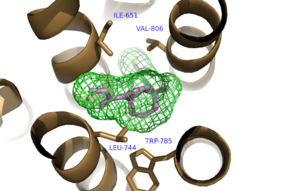
[[Image:Mav_Hydrophobic_pocket.png |300 px|left|thumb|Figure 1. Hydrophobic Pocket Surrounding Mavoglurant]] | [[Image:Mav_Hydrophobic_pocket.png |300 px|left|thumb|Figure 1. Hydrophobic Pocket Surrounding Mavoglurant]] | ||
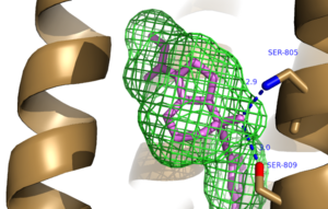
| - | [[Image:Mav_HB_2.png|300 px|left|thumb|Figure 2. Hydrogen Bonding between mGlu<sub>5</sub> and Mavoglurant. Blue coloration represents Hydrogen Bond | + | [[Image:Mav_HB_2.png|300 px|left|thumb|Figure 2. Hydrogen Bonding between mGlu<sub>5</sub> and Mavoglurant. Blue coloration represents Hydrogen Bond donor whereas red coloration represents Hydrogen Bond acceptor.]] |
</StructureSection> | </StructureSection> | ||
== References == | == References == | ||
<references/> | <references/> | ||
Revision as of 17:20, 16 April 2016
| |||||||||||
References
- ↑ Vassilatis DK, Hohmann JG, Zeng H, Li F, Ranchalis JE, Mortrud MT, Brown A, Rodriguez SS, Weller JR, Wright AC, Bergmann JE, Gaitanaris GA. The G protein-coupled receptor repertoires of human and mouse. Proc Natl Acad Sci U S A. 2003 Apr 15;100(8):4903-8. Epub 2003 Apr 4. PMID:12679517 doi:http://dx.doi.org/10.1073/pnas.0230374100
- ↑ 2.0 2.1 Venkatakrishnan AJ, Deupi X, Lebon G, Tate CG, Schertler GF, Babu MM. Molecular signatures of G-protein-coupled receptors. Nature. 2013 Feb 14;494(7436):185-94. doi: 10.1038/nature11896. PMID:23407534 doi:http://dx.doi.org/10.1038/nature11896
- ↑ Pin JP, Galvez T, Prezeau L. Evolution, structure, and activation mechanism of family 3/C G-protein-coupled receptors. Pharmacol Ther. 2003 Jun;98(3):325-54. PMID:12782243
- ↑ 4.0 4.1 4.2 4.3 4.4 4.5 4.6 4.7 4.8 Dore AS, Okrasa K, Patel JC, Serrano-Vega M, Bennett K, Cooke RM, Errey JC, Jazayeri A, Khan S, Tehan B, Weir M, Wiggin GR, Marshall FH. Structure of class C GPCR metabotropic glutamate receptor 5 transmembrane domain. Nature. 2014 Jul 31;511(7511):557-62. doi: 10.1038/nature13396. Epub 2014 Jul 6. PMID:25042998 doi:http://dx.doi.org/10.1038/nature13396
- ↑ Shigemoto R, Nomura S, Ohishi H, Sugihara H, Nakanishi S, Mizuno N. Immunohistochemical localization of a metabotropic glutamate receptor, mGluR5, in the rat brain. Neurosci Lett. 1993 Nov 26;163(1):53-7. PMID:8295733
- ↑ Li G, Jorgensen M, Campbell BM. Metabotropic glutamate receptor 5-negative allosteric modulators for the treatment of psychiatric and neurological disorders (2009-July 2013). Pharm Pat Anal. 2013 Nov;2(6):767-802. doi: 10.4155/ppa.13.58. PMID:24237242 doi:http://dx.doi.org/10.4155/ppa.13.58


