User:Camille Zumstein/Sandbox
From Proteopedia
(Difference between revisions)
| Line 16: | Line 16: | ||
<scene name='75/750223/Jw_caln_sec_strcutur/1'>Calcineurin</scene> is a heterodimeric Protein that consits of two subunits. They are called catalytic and regulatory subunit. | <scene name='75/750223/Jw_caln_sec_strcutur/1'>Calcineurin</scene> is a heterodimeric Protein that consits of two subunits. They are called catalytic and regulatory subunit. | ||
| - | This structure presented in this article is the catalytic subunit isoform of the serine/threonine-protein phosphatase 2B in rattus norvegicus (rat). It consists of 521 <ref>http://www.uniprot.org/uniprot/P63329</ref> aminoacids and has a molecular weight of 57 kDa. | + | This structure presented in this article is the catalytic subunit isoform of the serine/threonine-protein phosphatase 2B in rattus norvegicus (rat). It consists of 521 <ref>http://www.uniprot.org/uniprot/P63329</ref> aminoacids and has a molecular weight of 57 kDa <ref>http://www.uniprot.org/uniprot/P63329</ref>. |
| + | The calatytic subunit is subdivided into functional domains which are a catalytic domain, a binding domain for the regulary subsunit, a calmodulin binding domain and an autoinhibitory domain. | ||
| - | '''Discovery of the | + | '''Discovery of the structure:''' |
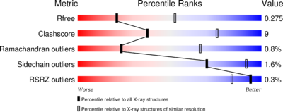
The structure have been published in the year 2013 by Qilu Ye et al. <ref>http://www.sciencedirect.com/science/article/pii/S0898656813002702</ref>. For their experiments they used [https://en.wikipedia.org/wiki/X-ray_crystallography#X-ray_diffraction x-ray diffraction]. The PDB validation obtained a Resolutionof 3.0 Å, a free R-value of 0.273 and a work R-value of 0.241 <ref>http://www.rcsb.org/pdb/explore/explore.do?structureId=4IL1</ref>. | The structure have been published in the year 2013 by Qilu Ye et al. <ref>http://www.sciencedirect.com/science/article/pii/S0898656813002702</ref>. For their experiments they used [https://en.wikipedia.org/wiki/X-ray_crystallography#X-ray_diffraction x-ray diffraction]. The PDB validation obtained a Resolutionof 3.0 Å, a free R-value of 0.273 and a work R-value of 0.241 <ref>http://www.rcsb.org/pdb/explore/explore.do?structureId=4IL1</ref>. | ||
[[Image:4il1 multipercentile validation.png|thumb|left|400 px]] | [[Image:4il1 multipercentile validation.png|thumb|left|400 px]] | ||
Revision as of 11:10, 8 January 2017
Structure Rat Calcineurin
| |||||||||||
References
- ↑ Hanson, R. M., Prilusky, J., Renjian, Z., Nakane, T. and Sussman, J. L. (2013), JSmol and the Next-Generation Web-Based Representation of 3D Molecular Structure as Applied to Proteopedia. Isr. J. Chem., 53:207-216. doi:http://dx.doi.org/10.1002/ijch.201300024
- ↑ Herraez A. Biomolecules in the computer: Jmol to the rescue. Biochem Mol Biol Educ. 2006 Jul;34(4):255-61. doi: 10.1002/bmb.2006.494034042644. PMID:21638687 doi:10.1002/bmb.2006.494034042644
- ↑ http://www.jimmunol.org/content/177/4/2681.full
- ↑ http://www.uniprot.org/uniprot/P63329
- ↑ http://www.uniprot.org/uniprot/P63329
- ↑ http://www.sciencedirect.com/science/article/pii/S0898656813002702
- ↑ http://www.rcsb.org/pdb/explore/explore.do?structureId=4IL1
- ↑ https://www.ncbi.nlm.nih.gov/pubmed/8811062
- ↑ http://www.uptodate.com/contents/pharmacology-of-cyclosporine-and-tacrolimus


