Grb2 protein: Growth Factor Receptor Bound Protein is a cytosolic protein made of 217 amino acids and weighing 25,206 Da. Ubiquitously present in the cell, this protein is involved in signal transduction and especially in the MAP kinase pathway. Grb2 interacts mainly with tyrosine kinase such as //en.wikipedia.org/w/api.php?action=query&prop=links&pllimit=500&titles=fullpagename your label once it’s been activated by ligand binding. This specific binding leads to the recruitment of GEF (like SOS1), stimulating the activation of other pathways. Several others interactions have been elucidated like the capacity of the protein to dimerise thus implicated in the growth of malignant cells.
Structure
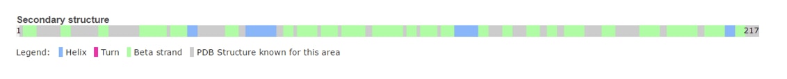
Grb2 protein has a very well characterized structure. Composed of 217 amino acids organized in two chains structured in B sheets and alpha helices.
 The protein has three main domains:
-
-
-
:
SH2 domain is a domain that is approximately 100 amino acids long and with a very conserved structure. Identified in in several human and rodent proteins such as phosphatases, TF, or adaptor like protein such as Grb2 for instance. This domain is therefore ubiquitous in several cellular signaling pathways.Typically, SH2 domain specifically recognize sites with phosphorylated tyrosine in different types of proteins. SH2 can for instance bind to the intracellular region of EGF leading in turn, to the formation of protein signalization complexes. This binding and the role of SH2 is thus, very important in the conversion of an extra-cellular signal in an intra-cellular signal able to give rise to diversified cellular responses or the expression of specific genes.It is also important to note that the SH2 domain can bind to other SH2 domains.However, a mutation in the specific binding site of SH2 can impede the interaction of two proteins and thus the formation of a protein complex. Therefore, mutations in SH2 can give rise to cellular dysfunction and lead to several diseases.
:
The SH3 domain is a region of a protein that is approximately 50 amino acid long. Largely present in proteins associated to the membrane. The domain is made of 5 to 6 Beta-sheets arranged in two antiparallel Beta-sheets. The linking region between the two Beta-sheets can contain alpha helices. This special conformation allows the arrangement of a hydrophobic pocket in which the ligand can bind. Typically, the binding region has a motif rich in Prolines: PXXP. This binding allows the formation of multi-proteins complexes involved in the translation of an extra-cellular signal and its conversion. The binding can thus be largely involved in gene expression and protein concentration.
ISOFORM:
Nevertheless, Grb2’s isoform (Grb3.3) is also present in the cell and induces apoptosis. This isoform has a very similar structure to Grb2 but is truncated from an SH3 domain (from the 60th amino acid to the 100th ) resulting in a degradation of its SH2 domain and therefore in a loss of functionality.
Function
The Grb2 isoform has a non-functional SH2 domain, unable to link the phosphorylated tyrosine of its targeted protein (EGFR for instance). This inability of the molecule to transmit signal is traduce by apoptosis of the cell, thus regulating the growth signal.
The functional isoform Grb2 is involved in several cellular functions detailed below. On one hand, the SH2 domain recognizes phosphorylated residues which are mainly tyrosines. The recognized tyrosines present a caracteristic motif for recognition : NH2-pYXNX-COOH.
   -	pY representing the phosphorylated tyrosine.
   -	N for Asparagine
   -	X for a random residue
Thus by the special recognition of this motif, the binding of the 2 molecules is very specific. These motifs are highly expressed in several cellular proteins like Receptor Tyrosine Kinase (epidermal growth factor receptor, fibroblast growth factor receptor, nerve growth factor receptor) but equally in proteins that are not RTK kinases (BCR-Ab1, focal adhesion kinase, insulin receptor substrate-1).
As an example, the SH2 domain of Grb2 recognizes an intracellular phosphorylated tyrosine. This binding, in turn, leads to the recruitment of SOS-1 via the SH3 domain of Grb2. Indeed, Grb2 is also made of two SH3 domains. These domains are able to recognize Proline rich region like the one of SOS-1 protein (Son Of Sevenless).
Following this pathway and the formation of a complex between Grb2 and SOS, the RAS protein is activated. Interestingly, RAS is a g-protein implicated in the activation of Raf-1. The latest activates of the MEK downstream cascade pathway (MEK1/ MEK2 et ERK1/ ERK2) involved in the translocation of ERF factors from the cytosol to the nucleus for the activation of Elk-1 and Myc transcription Factor (TF). These particular TF participate in the activation of SRE containing gene leading to cellular growth.
On the other hand, in T lymphocytes, the simulation of TCRs induces tyrosine phosphorylation on a wide range of of cellular proteins such as p36-38 or LAT.
As an example, the phosphorylated residues of LAT can bind the SH2 domain of Grb2 while the formation of this complex recruits on the SH3 domain some proteins of the VAV family. VAV proteins are guanine nucleotide exchange factors (GEF) for the GTPase proteins of the Rho family.
This complex has for main aim to introduce a Calcium flux and the activation of MAP kinase allowing lymphocytes T proliferation.
Finally, it was proven that Grb2 in the negative regulation of EGFR. Indeed, c-Cbl is a protein implicated in the E3 complex of EGFR ubiquitination, hence also its degradation. C-Cbl thanks to its SH2 domain can directly bind to EGFR causing its degradation (Grb2 independent regulation). Yet c-Cbl can also indirectly bind to EGFR via its SH3 domain recognition by Grb2 (Dependant Grb2 regulation). The direct or indirect binding of c-Cbl on EGFR induce the recruitment of enzymes that are necessary for the ubiquitination of EGFR. Ubiquitination being a signal for protein degradation. It is important to note that negative regulation is more important when Grb2 is implicated and bound to c-Cbl rather than when c-Cbl is the only protein involved.
Interactions
Sos1: Promotes the exchange of Ras-bound GDP into GTP, by promoting the Ras specific guanine nucleotide exchange factor activity.
Shc: Shc is important in the regulation of apoptosis and drug resistance in mammalian cells. She is implicated in the EGFR pathway.
Cbl: Cbl is a pronto oncogene protein which serves as an adaptor and a negative regulator of many signalling pathways implicated in cell surface receptors activation.
Gab2: Gab2 acts downstream of several membrane receptors such as cytokine, hormone, cell matrix or growth factor receptor. Thus, it is implicated in many different pathways.
LCP2: Involved in T cell antigen receptor mediated signaling
Erbb2: Erbb2 is a kinase involved in several surface receptor complexes, but need a co receptor for ligand binding. For example, it participates in neuregulin receptor complex but it can’t bind with it alone.
Frs2: Fibroblast growth factor receptor substrate 2 can link to FGR and NGF activated receptor. They play an important role in the activation of MAPK kinase for example, or the phosphorylation of PIK3R1.
Irs1: Insulin receptor substrate 1 may mediate the control of various cellular processes by insulin. It can activate the phosphatidylinositol 3 kinase when it bounds to the regulatory p85 subunit.
Gab1: GRB2 associated binding protein 1, is implicated in many signalling cascades triggered by activated receptor type kinases. It is also probably involved in signalling by the epidermal growth factor receptor.
EGFR: The epidermal growth factor receptor has a Tyrosine kinase activity and can be recognize by Grb2 thanks to its Tyrosine domains. This receptor is implicated in many pathways, such as antigen fixation on B cells.
EGFR interaction
Disease
Alzheimer’s Disease (AD): It seems like Grb2 is implicated in the simulation of AD. Phenotypic change have been identified in cortical and hippocampal neurons characteristic of AD. Indeed, the proteins implicated in the transduction of the signal from Grb2 to SOS are altered in AD. This modifications would be at the heart at the transduction of a “derived” signal stimulating AD. [1]
HIV-1: Grb2 isoform could have a simulatory effect in the retro viral infection of HIV-1. By its essential role in the MAPK pathway, Grb3 can have effects on HIV-1 infections. Indeed, the replication of the virus is activated by Lymphocytes T replication. Yet lymphocytes T’s activation depend on the activation of the MAPK pathway dictated by the presence or not of grb3 in the cell. This pathway finally activates NFAT TF, a TF enhancing the LTR promotor of HIV-1 leading to its replication.
Relevance
Grb2 protein is especially involved in the setting up of cellular oncognesis in prostate, colon and lung cancers. This role is mainly due to its essential role in signal transduction in the MAP kinase pathway known to induce mitosis.
In this pathway, GrbS binds to the oncogenic protein SOS under its monomeric form. Yet SOS can also be found in its dimeric form in the cell.
Dimerization of Grb2 is dependent upon several factors like the phosphorylation of or the binding of ligand on the SH2 domain of the same protein. Mainly, phosphorylation induce the dissociation of the Grb2 dimer induce an increase in the MAP kinase pathway activation by the binding of SOS. The phosphorylated state of has been discovered in severa pre-metastatis cancers. This highly suggest that pY160 could be a oncogenic marker in humans. A new therapeutic way could therefore be considered by stabilizing Grb2 in its dimeric form. This could be achieve with a protein acting as an irreversible cross-link at the interface between the 2 units.
References
- ↑ McShea A, Zelasko DA, Gerst JL, Smith MA. Signal transduction abnormalities in Alzheimer's disease: evidence of a pathogenic stimuli. Brain Res. 1999 Jan 9;815(2):237-42. PMID:9878757


