We apologize for Proteopedia being slow to respond. For the past two years, a new implementation of Proteopedia has been being built. Soon, it will replace this 18-year old system. All existing content will be moved to the new system at a date that will be announced here.
Image:3h90sections.PNG
From Proteopedia
(Difference between revisions)
No higher resolution available.
3h90sections.PNG (423 × 527 pixel, file size: 101 KB, MIME type: image/png)
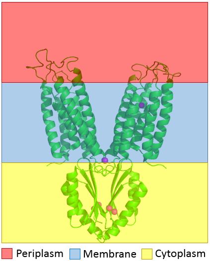
(The distribution of YiiP through the membrane is shown. The CTD is shown in highlighted in the yellow bottom to show that it is in the cytoplasm and the TMD is highlighted with blue to show that it sits in membrane.) |
|||
| (One intermediate revision not shown.) | |||
| Line 1: | Line 1: | ||
| - | The distribution of YiiP through the membrane is shown. The CTD is shown | + | The distribution of YiiP through the membrane is shown. The CTD is shown highlighted in the yellow to show that it is in the cytoplasm and the TMD is highlighted with blue to show that it sits in the membrane. |
Current revision
The distribution of YiiP through the membrane is shown. The CTD is shown highlighted in the yellow to show that it is in the cytoplasm and the TMD is highlighted with blue to show that it sits in the membrane.
File history
Click on a date/time to view the file as it appeared at that time.
| Date/Time | User | Dimensions | File size | Comment | |
|---|---|---|---|---|---|
| (current) | 18:05, 21 April 2017 | Kyle Colston (Talk | contribs) | 423×527 | 101 KB | The distribution of YiiP through the membrane is shown. The CTD is shown in highlighted in the yellow bottom to show that it is in the cytoplasm and the TMD is highlighted with blue to show that it sits in membrane. |
- Edit this file using an external application
See the setup instructions for more information.
Links
The following pages link to this file:
