Thiolase
From Proteopedia
(Difference between revisions)
| Line 8: | Line 8: | ||
== Structural highlights == | == Structural highlights == | ||
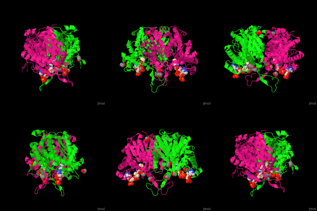
| - | The <scene name=' | + | The <scene name='44/447919/Cv/2'>active site of 3-ketoacyl-CoA thiolase contains CoA</scene><ref>PMID:25478839</ref>. Water molecules shown as red spheres. |
</StructureSection> | </StructureSection> | ||
== 3D Structures of Thiolase == | == 3D Structures of Thiolase == | ||
Revision as of 11:39, 9 October 2017
| |||||||||||
3D Structures of Thiolase
Updated on 09-October-2017
References
- ↑ Sundaramoorthy R, Micossi E, Alphey MS, Germain V, Bryce JH, Smith SM, Leonard GA, Hunter WN. The crystal structure of a plant 3-ketoacyl-CoA thiolase reveals the potential for redox control of peroxisomal fatty acid beta-oxidation. J Mol Biol. 2006 Jun 2;359(2):347-57. Epub 2006 Mar 29. PMID:16630629 doi:http://dx.doi.org/10.1016/j.jmb.2006.03.032
- ↑ Soto G, Stritzler M, Lisi C, Alleva K, Pagano ME, Ardila F, Mozzicafreddo M, Cuccioloni M, Angeletti M, Ayub ND. Acetoacetyl-CoA thiolase regulates the mevalonate pathway during abiotic stress adaptation. J Exp Bot. 2011 Nov;62(15):5699-711. doi: 10.1093/jxb/err287. Epub 2011 Sep 9. PMID:21908473 doi:http://dx.doi.org/10.1093/jxb/err287
- ↑ Kiema TR, Harijan RK, Strozyk M, Fukao T, Alexson SE, Wierenga RK. The crystal structure of human mitochondrial 3-ketoacyl-CoA thiolase (T1): insight into the reaction mechanism of its thiolase and thioesterase activities. Acta Crystallogr D Biol Crystallogr. 2014 Dec 1;70(Pt 12):3212-25. doi:, 10.1107/S1399004714023827. Epub 2014 Nov 22. PMID:25478839 doi:http://dx.doi.org/10.1107/S1399004714023827

