Biotin Protein Ligase
From Proteopedia
(Difference between revisions)
| Line 7: | Line 7: | ||
Biotinylation is catalysed through a two-step reaction where biotin is first activated to biotinyl-5′-AMP in an ATP dependent manner. The biotin is then transferred onto the ε-amino group of a specific target lysine residue. The reaction mechanism is related to that of amino acyl-tRNA synthetases and lipoyl ligases where the reaction proceeds through the formation of an adenylated intermediate, suggesting a common ancestral relationship <ref>pmid 18442489</ref> . | Biotinylation is catalysed through a two-step reaction where biotin is first activated to biotinyl-5′-AMP in an ATP dependent manner. The biotin is then transferred onto the ε-amino group of a specific target lysine residue. The reaction mechanism is related to that of amino acyl-tRNA synthetases and lipoyl ligases where the reaction proceeds through the formation of an adenylated intermediate, suggesting a common ancestral relationship <ref>pmid 18442489</ref> . | ||
| - | Of all the BPL’s, | + | Of all the BPL’s, '''BirA bifunctional protein''' is by far the most characterised and understood family member. BirA catalyzes the two activities of post-translational biotinylation and repression of transcription initiation<ref>pmid 28466579</ref>. A recent ensemble of BPL structures from the thermophilic archea Pirococcus Horikoshii OT3 <ref>pmid 16510991</ref> have also provided new insights into the catalytic mechanism of BPLs. |
== Structural highlights == | == Structural highlights == | ||
| Line 31: | Line 31: | ||
**[[3fjp]], [[2eay]] – AaBPL – ''Aquifex aeolicu''s<br /> | **[[3fjp]], [[2eay]] – AaBPL – ''Aquifex aeolicu''s<br /> | ||
**[[3efr]] – AaBPL (mutant) <br /> | **[[3efr]] – AaBPL (mutant) <br /> | ||
| - | **[[1bia]] – EcBPL – ''Escherichia coli'' <br /> | ||
**[[3rkx]], [[3v8j]] – SaBPL – ''Staphylococcus aureus'' | **[[3rkx]], [[3v8j]] – SaBPL – ''Staphylococcus aureus'' | ||
| Line 50: | Line 49: | ||
**[[4ha8]] - SaBPL + biotin derivative<br /> | **[[4ha8]] - SaBPL + biotin derivative<br /> | ||
**[[3v7s]], [[3v7r]] – SaBPL + inhibitor<br /> | **[[3v7s]], [[3v7r]] – SaBPL + inhibitor<br /> | ||
| - | **[[1bib]], [[1hxd]] - EcBPL + biotin<br /> | ||
| - | **[[2ewn]] - EcBPL + biotinyl-AMP<br /> | ||
| - | **[[4wf2]] - EcBPL (mutant) + biotinyl-AMP<br /> | ||
*Biotin protein ligase ternary complex | *Biotin protein ligase ternary complex | ||
| Line 60: | Line 56: | ||
**[[2dto]], [[2dth]], [[2fyk]] - PhBPL + ATP + biotin<br /> | **[[2dto]], [[2dth]], [[2fyk]] - PhBPL + ATP + biotin<br /> | ||
**[[2dkg]] - PhBPL + biotinyl-AMP + pyrophosphate | **[[2dkg]] - PhBPL + biotinyl-AMP + pyrophosphate | ||
| + | |||
| + | *BirA bifunctional protein | ||
| + | |||
| + | **[[1bia]] – EcBirA – ''Escherichia coli''<br /> | ||
| + | **[[1bib]], [[1hxd]] - EcBirA + biotin<br /> | ||
| + | **[[2ewn]] - EcBirA + biotinyl-AMP<br /> | ||
| + | **[[4wf2]] - EcBirA (mutant) + biotinyl-AMP<br /> | ||
| + | **[[3rux]], [[4xtu]], [[4xtv]], [[4xtw]], [[4xtx]], [[4xty]], [[4xtz]], [[4xu0]], [[4xu1]], [[4xu2]], [[4xu3]] - MtBirA + inhibitor<br /> | ||
| + | **[[4op0]] - MtBirA + biotinyl-AMP<br /> | ||
| + | **[[6apw]], [[6aqq]] - SaBirA + inhibitor<br /> | ||
| + | |||
}} | }} | ||
==References== | ==References== | ||
Revision as of 10:10, 5 March 2018
| |||||||||||
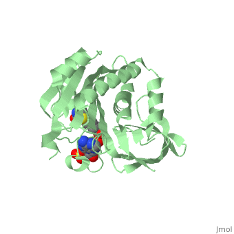
3D structures of Biotin Protein Ligase
Updated on 05-March-2018
References
- ↑ Pendini NR, Bailey LM, Booker GW, Wilce MC, Wallace JC, Polyak SW. Microbial biotin protein ligases aid in understanding holocarboxylase synthetase deficiency. Biochim Biophys Acta. 2008 Jul-Aug;1784(7-8):973-82. Epub 2008 Apr 9. PMID:18442489 doi:10.1016/j.bbapap.2008.03.011
- ↑ Wang J, Beckett D. A conserved regulatory mechanism in bifunctional biotin protein ligases. Protein Sci. 2017 Aug;26(8):1564-1573. doi: 10.1002/pro.3182. Epub 2017 May 11. PMID:28466579 doi:http://dx.doi.org/10.1002/pro.3182
- ↑ Bagautdinov B, Kuroishi C, Sugahara M, Kunishima N. Purification, crystallization and preliminary crystallographic analysis of the biotin-protein ligase from Pyrococcus horikoshii OT3. Acta Crystallogr Sect F Struct Biol Cryst Commun. 2005 Feb 1;61(Pt, 2):193-5. Epub 2005 Jan 8. PMID:16510991 doi:10.1107/S1744309104034360
- ↑ Wilson KP, Shewchuk LM, Brennan RG, Otsuka AJ, Matthews BW. Escherichia coli biotin holoenzyme synthetase/bio repressor crystal structure delineates the biotin- and DNA-binding domains. Proc Natl Acad Sci U S A. 1992 Oct 1;89(19):9257-61. PMID:1409631
Proteopedia Page Contributors and Editors (what is this?)
Michal Harel, Nicole R Pendini, Alexander Berchansky, David Canner, Jaime Prilusky
