This old version of Proteopedia is provided for student assignments while the new version is undergoing repairs. Content and edits done in this old version of Proteopedia after March 1, 2026 will eventually be lost when it is retired in about June of 2026.
Apply for new accounts at the new Proteopedia. Your logins will work in both the old and new versions.
User:Mark Hoelzer/Sandbox1
From Proteopedia
(Difference between revisions)
| Line 6: | Line 6: | ||
<table> | <table> | ||
<tr> | <tr> | ||
| - | <td> | + | <td><StructureSection load='1a3n' size='400' caption='Hemoglobin based on 1a3n.pdb' scene=''></td> |
| - | <td> | + | <td>[[Image:Cbm_hemoglobin1.jpg]]</td> |
</tr> | </tr> | ||
</table> | </table> | ||
| - | {| class="wikitable" | ||
| - | |- | ||
| - | | <StructureSection load='1a3n' size='400' caption='Hemoglobin based on 1a3n.pdb' scene=''> | ||
| - | | [[Image:Cbm_hemoglobin1.jpg | left]] | ||
| - | |} | ||
Revision as of 14:45, 3 April 2018
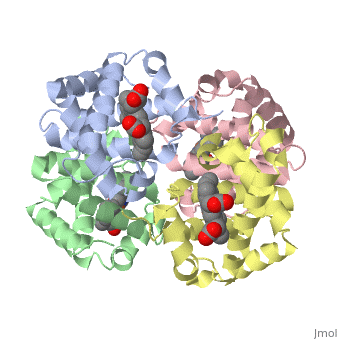
3D Printed Physical Model of Hemoglobin
Shown below is a physical 3D printed model of Hemoglobin, based on the structure 1a3n.pdb. The two alpha-globin chains are colored light red, the two beta globin chains are colored dark red, and the four heme groups are colored yellow.
|
||||||||||||