Overview
DNA polymerases are enzymes that play a key role in DNA replication. DNA replication is the process of splitting an existing double-stranded DNA molecule into two single strands of DNA, then using DNA polymerases to translate the single strands. The process of translation results in the creation of the complementary DNA strands and results in the creation of two double-stranded DNA molecules that are exact replicas of the original DNA molecule. The complementary strands are created in the 5'-3' direction. Certain DNA polymerases are also responsible for proofreading the newly synthesized DNA strand and using exonuclease to remove and replace any errors that occurred. DNA polymerases are divided into 7 families according to their sequence homology and 3D structure similarities.[1] The families are:
- Family A - DNA replication and repair (includes DNA Polymerase I, γ)
- Family B - DNA replication and repair (includes DNA Polymerase II, α, δ, ε)
- Family C - DNA replication in prokaryotes (includes DNA Polymerase III)
- Family D - DNA replication in archaea
- Family X - DNA repair in eukaryotes (includes DNA Polymerase β, λ, μ)
- Family Y - DNA replication of damaged DNA (includes DNA Polymerase IV, V, η, ι, κ)
- Family RT - reverse transcriptase (See Reverse transcriptase.)
Function
DNA polymerases are essential enzymes for DNA Replication.[1] Before DNA polymerases can perform its part in DNA replication, other enzymes must unwind and split the double helical structure of DNA and signal for the initiation of replication. Once DNA primase has placed a primer on the template DNA strand, DNA polymerases can attach. These enzymes use the template strand of DNA to synthesize a complementary strand of DNA using the DNA building blocks called nucleotides. The order of the nucleotides on the complementary strand is determined by the base-pairing rules: cytosine with guanine and adenine with thymine.
During DNA synthesis, the DNA polymerases move along the template DNA strand in a 3'-5' direction and adds nucleotides to the new DNA strand in a 5'-3' direction. This causes the elongation of the new strand in a 5'-3' direction. Note that the direction of the newly formed DNA strand is opposite of the template DNA strand. This makes the resulting double-stranded DNA molecule complementary and anti-parallel.
DNA polymerases are some of the most accurate enzymes and have about one mistake for every one billion copies. When a mistake is made, many of the DNA polymerases have the ability to proofread the newly synthesized DNA and correct any mistakes made during replication. The enzymes proofread in the 5'-3' direction. When an error is found, the misplaced nucleotide is cut out so the correct nucleotide can be inserted. This process is often referred to as 5'-3'exonuclease activity.
Types of DNA Polymerase
According to their sequence homology and 3D structure similarities, DNA Polymerases can be divided into 7 families: A, B, C, D, X, Y, and RT.[2]
| Family
| Function
| Species
| Examples
|
| A
| Replication and Repair
| Eukaryotes and Prokaryotes
| Pol I and Pol γ
|
| B
| Replication and Repair
| Eukaryotes and Prokaryotes
| Pol II, Pol α, Pol δ, and Pol ε
|
| C
| Replication
| Prokaryotes
| Pol III
|
| D
| Replication
| Archaea
| Unknown
|
| X
| Replication and Repair
| Eukaryotes
| Pol β, Pol μ, and Pol λ
|
| Y
| Replication and Repair
| Eukaryotes and Prokaryotes
| Pol IV, Pol V, Pol η, Pol κ, and Pol ι
|
| RT
| Replication and Repair
| Eukaryotes, Viruses, and Retrovirus
| Telomerase and Hepatitis B virus
|
Eukaryotic Polymerase
Polymerase γ
Polymerase γ is considered a Family A polymerase. Pol γ's main function is to replicate and repair mitochondrial DNA (mtDNA). Pol γ can perform proofreading 3'–5' exonuclease activity. Mutations that cause limited or non-functioning Pol γ has a significant effect on mtDNA and is a common cause of autosomal mitochondrial disorders.[3]
Polymerase α, Polymerase δ, and Polymerase ε
Members of family B, Pol α, Pol δ, and Pol ε are the main polymerases involved in DNA replication. Pol α binds with primase to form a complex. Primase creates and places an RNA primer, allowing Pol α to start replication. Pol δ then takes over the synthesis of the lagging strand from Pol α. It is believed that Pol ε synthesizes the leading strand during replication, while Pol δ primarily replicates the lagging strand. However, there have been some cases where Pol δ has been found to replicate the lagging and leading strand. Pol δ and ε also possess 3'-5' exonuclease activity capabilities.[3]
Family X
Family X polymerases consist of polymerases like Pol β, Pol μ, and Pol λ. Pol β's main function is short-patch base excision repair, a repair pathway used for repairing alkylated or oxidized bases. Pol λ and Pol μ are essential for rejoining DNA double-strand breaks due to hydrogen peroxide and ionizing radiation, respectively.[3] For more details see DNA polymerase beta.
Polymerases η, Polymerase ι, and Polymerase κ
Polymerase η, Polymerase ι, and Polymerase κ are Family Y DNA polymerases involved in the DNA repair by translesion synthesis. Polymerases in Family Y are prone to errors during DNA synthesis. Pol η is important for the accurate translesion synthesis of DNA damage resulting from ultraviolet radiation. The function of Pol κ is not completely understood, but it is thought to act as an extender or inserter of a specific base at certain DNA lesions. All three translesion synthesis polymerases are activated by stalled replicative DNA polymerases.[3]
Prokaryotic Polymerase
DNA Polymerase I
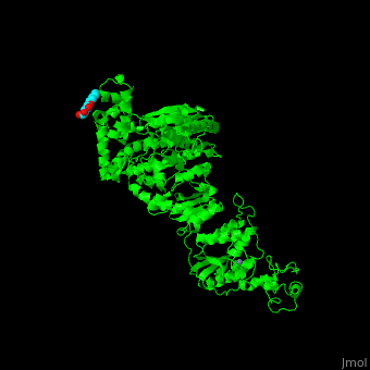
DNA Polymerase I is a family A enzyme whose main function is excision repair of DNA strands through 3'-5' and 5'-3' exonuclease. This polymerase also helps with Okazaki fragment maturation. Okazaki fragments are short synthesized strands of DNA that form the lagging strand during DNA replication. When Polymerase I does replicate, it starts adding nucleotides at the RNA primer and moves in the 5'-3' direction. This polymerase is also the major polymerase in E. coli.[3] See also Taq DNA polymerase (Hebrew).
in Family A DNA polymerase I (1taq).
in Family A DNA polymerase I (1taq).[4]
DNA Polymerase II
DNA polymerase II belongs to family B. It is responsible for 3'-5' exonuclease activity and restarting replication after the synthesis process has stopped due to damage in the DNA strand. Polymerase II is located at the replication fork in order to help direct the activity of other polymerases.[3]
DNA Polymerase III
DNA polymerase III is the primary enzyme involved in the replication of DNA. It belongs to family C and is responsible for synthesizing new DNA strands by adding nucleotides to the 3'-OH group of the primer. This enzyme also has 3'-5' exonuclease activity giving it the ability to check the synthesized DNA strand for errors.[3]
For more details see Polymerase III homoenzyme beta subunit and Alpha Subunit of Thermus aquaticus DNA Polymerase III.
DNA Polymerase IV
DNA polymerase IV is involved in non-targeted mutagenesis. Belonging to family Y, this enzyme is activated when synthesis at the replication fork stalls. once activated, Polymerase IV creates a checkpoint, stops replication, and allows time to properly repair lesions in the DNA strand. Polymerase IV is also involved in translesion synthesis, a DNA repair mechanism. However, the enzyme lacks nuclease activity making it prone to errors in DNA replication.[3]
DNA Polymerase V
DNA polymerase V, in family Y, is highly regulated and only produced when DNA is damaged and requires translesion synthesis. Polymerase V, like polymerase IV, lacks all exonuclease function and is unable to proofread the synthesized DNA strand causing it to be less efficient.[3]
Reverse Transcriptase
See Reverse transcriptase.
Structure
The basic structure of all DNA polymerases consists of subdomains referred to as the palm, fingers, and thumb. The palm contains catalytically essential amino acids in it's active sites. The fingers are essential for nucleotide recognition and binding. The thumb is important for the binding of the DNA substrate. These subdomains, along with other subdomains specific to each family, are essential for the correct functioning of DNA polymerase. The structures of each of these subdomains are slightly different for each polymerase, but not much is known about those subtle differences. [5]
Family A
In addition to the basic structure of DNA polymerase, the Family A polymerases also have a 5'-3' exonuclease that is required for the removal of RNA primers from Okazaki fragments. Not all, but some Family A polymerases also a 3'-5' exonuclease that is responsible for proofreading the DNA. [5]
Family B
In addition to the basic structure of DNA polymerase, the Family B polymerases contain an extremely active 3'-5' exonuclease that corrects errors in DNA replication. [5]
Family X
The thumb, palm, and fingers subdomains are a part of the of N-terminal, or 31-kDA polymerase fragment in the Family X Polymerases. The palm in this family contains three aspartic acid motifs. The fingers in this family have Helices M and N that contain amino acid residues. [6] The N-terminal is connected to an 8kDa amino terminal domain containing a 5' deoxyribose phosphate lyase that is required for base excision repair. Each member contains it's own structural differences that aid in it's functioning. [5]
Family Y
The N-terminal of the Family Y polymerases contains the catalytic core of the fingers, palm, and thumb. The C-terminal, which has a conserved tertiary structure of a four-stranded beta sheet supported on one side by two alpha helices, otherwise referred to as the little finger domain, contributes to DNA binding and is essential for complete polymerase activity. This family lacks flexibility in the fingers subdomain, which is uncharacteristic of the other families. The other parts of the catalytic core and the little finger domain are flexible and frequently assume different positions. [7]
Mechanism
The majority of DNA polymerases undergo a two-metal-ion mechanism. Two metal ions in the active site work to stabilize the pentacoordinated transition state. The first metal ion activates the hydroxyl groups. Those hydroxyl groups then go on to attack the phosphate group of the dNTP. The second metal ion not only stabilizes the negative charge, but also builds on the leaving oxygen and chelating phosphate groups. [8]
Some Dpo terminology:
Dpo sliding clamp is made of the complex of Dpo and Proliferating Cell Nuclear Antigen (PCNA) which encircles it.
The BRCT domain in Dpo is the C-terminal domain of breast cancer susceptibility protein.
Klenow fragment is a large Dpo fragment produced upon cleavage of Dpo by subtilisin.
In the E. coli, the EcDpo III subunits β, γ, δ, δ' are named clamp loader. This complex assembles the β subunit sliding clamp unto the DNA.
See also User:Karl E. Zahn/RB69 DNA polymerase (gp43)