User:Karsten Theis/RNaseA physical model explanation
From Proteopedia
(Difference between revisions)
(→Tour of what the model shows) |
|||
| Line 7: | Line 7: | ||
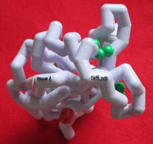
Mark Hoelzer from the MSOE Center for Biomolecular Modeling [http://cbm.msoe.edu/about/staff.php] designed, printed and painted the model. Having the model in your hand is great because you can experience the structure in a very direct way, and having this companion page is great because you can see the model even if you do not have the physical model in your hand. It also allows you to hover over different parts of the model with a mouse to get more information (try it after turning off spinning with the +/- spin button!). | Mark Hoelzer from the MSOE Center for Biomolecular Modeling [http://cbm.msoe.edu/about/staff.php] designed, printed and painted the model. Having the model in your hand is great because you can experience the structure in a very direct way, and having this companion page is great because you can see the model even if you do not have the physical model in your hand. It also allows you to hover over different parts of the model with a mouse to get more information (try it after turning off spinning with the +/- spin button!). | ||
| - | ==Tour of | + | ==Tour of the structure== |
<StructureSection load='' size='340' side='right' caption='Caption for this structure' scene='78/785360/Physmodel/1'> | <StructureSection load='' size='340' side='right' caption='Caption for this structure' scene='78/785360/Physmodel/1'> | ||
| + | |||
| + | ==What the model shows== | ||
===Trace of alpha carbon atoms=== | ===Trace of alpha carbon atoms=== | ||
| Line 19: | Line 21: | ||
===Hydrogen bonds=== | ===Hydrogen bonds=== | ||
| - | + | <jmol><jmolLink> | |
| + | <script> select all; color hbonds blue; delay 0.8; color hbonds ivory | ||
| + | </script> | ||
| + | <text>highlight hydrogen bonds</text> | ||
| + | </jmolLink></jmol> | ||
===N-terminus and C-terminus=== | ===N-terminus and C-terminus=== | ||
Revision as of 16:39, 9 May 2018
Introduction
This proteopedia page is intended as a companion to a physical model of RNase A used for teaching protein folding and structure at Westfield State University.
Mark Hoelzer from the MSOE Center for Biomolecular Modeling [1] designed, printed and painted the model. Having the model in your hand is great because you can experience the structure in a very direct way, and having this companion page is great because you can see the model even if you do not have the physical model in your hand. It also allows you to hover over different parts of the model with a mouse to get more information (try it after turning off spinning with the +/- spin button!).
Tour of the structure
| |||||||||||

