Dinoflagellate luciferase
From Proteopedia
(Difference between revisions)
m |
|||
| (15 intermediate revisions not shown.) | |||
| Line 1: | Line 1: | ||
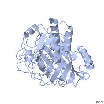
| - | + | <StructureSection load='1vpr' size='300' side='right' scene='' caption='Luciferase domain from a Dinoflagellate. Selenomethionines shown as space-filling objects [[1vpr]]'> | |
| - | + | ||
| - | + | ||
| - | + | ||
== Introduction == | == Introduction == | ||
| Line 10: | Line 7: | ||
== Structure == | == Structure == | ||
| - | Composed of residues 868-1218, domain 3 (D3) also consists of a 20aa C-terminal unresolved domain. Containing 7 α-helices and 16 β-strands, D3 is further organized into subdomains. The main portion of the enzyme appears to be a β-barrel structure composed of 10 anti-parallel strands connected via a Gly rich sequence to a 3 helix bundle. This bundle is stabilized by a hydrophobic core region as well as a multitude of H-bonding patterns<ref name="main" />. The β-barrel structure actually has some homology with the human muscle fatty acid binding protein (m-FABP, pdb= | + | Composed of residues 868-1218, domain 3 (D3) also consists of a 20aa C-terminal unresolved domain. Containing 7 α-helices and 16 β-strands, D3 is further organized into subdomains. The main portion of the enzyme appears to be a β-barrel structure composed of 10 anti-parallel strands connected via a Gly rich sequence to a 3 helix bundle. This bundle is stabilized by a hydrophobic core region as well as a multitude of H-bonding patterns<ref name="main" />. The β-barrel structure actually has some homology with the human muscle fatty acid binding protein (m-FABP, pdb= [[1hmt]]). Both are part of a "β-clam" subdomain family, responsible for binding of hydrophobic molecules. However, other known β-clam structures do not possess enzymatic activity<ref name="main" />. |
| Line 24: | Line 21: | ||
| - | [[Image:Luciferase_reaction.jpg]] | + | [[Image:Luciferase_reaction.jpg|left|thumb|450px]] |
''Image courtesy of L. Wayne Schultz.'' | ''Image courtesy of L. Wayne Schultz.'' | ||
| + | == References == | ||
| + | <references /> | ||
| + | ==Content Donors== | ||
| + | |||
| + | All the initial portions of this page were created by [[User:James Jones|James Jones]] and were moved because it deserved its own separate page distinct from the associated PDB entry. | ||
| + | </StructureSection> | ||
| + | ==3D structures of luciferase== | ||
| - | == Related Links == | ||
[[Luciferase]] | [[Luciferase]] | ||
| - | [[1vpr]] is the structure used on this page. | + | ==See Also== |
| + | *[[1vpr]] is the structure used on this page. | ||
| + | *[[Colored & Bioluminescent Proteins]] | ||
| + | * [[PyMOL]] | ||
| - | [http://www.pymol.org/ Pymol molecular viewer] | ||
| - | + | == External Resources == | |
| - | [http://www. | + | *[http://www.pdb.org/pdb/explore/explore.do?structureId=1VPR Protein Data Bank file on 1VPR] |
| - | [http:// | + | *[http://www.ncbi.nlm.nih.gov/protein/ABO61076.1?ordinalpos=1&itool=EntrezSystem2.PEntrez.Sequence.Sequence_ResultsPanel.Sequence_RVDocSum NCBI protein entry on ''P. lunula'' luciferase] |
| - | + | *[http://dx.doi.org/10.2210/rcsb_pdb/mom_2006_6 PDB molecule of the month feature on luciferases] | |
| - | + | [[Category:Topic Page]] | |
| - | + | ||
| - | + | ||
| - | + | ||
| - | + | ||
Current revision
| |||||||||||


3D structures of luciferase
See Also
- 1vpr is the structure used on this page.
- Colored & Bioluminescent Proteins
- PyMOL
External Resources
Proteopedia Page Contributors and Editors (what is this?)
Wayne Decatur, Michal Harel, Alexander Berchansky, James Jones, Joel L. Sussman, David Canner
