Main Page

From Proteopedia

(Difference between revisions)
Jump to: navigation, search
Line 62: Line 62:
<table width='100%' style="padding: 10px; background-color: #d7d8f9; font-size: 1.5em;"><tr>
<td>[[Proteopedia:About|About]]</td>
-
<td>[http://proteopedia.org/cgi-bin/contact Contact]</td>
+
<td>[[Special:Contact|Contact]]</td>
<td>[[Proteopedia:Table of Contents|Table of Contents]]</td>
<td>[[Proteopedia:Structure Index|Structure Index]]</td>

Revision as of 09:20, 21 October 2018

ISSN 2310-6301

As life is more than 2D, Proteopedia helps to bridge the 3D relationships between function & structure of biomacromolecules
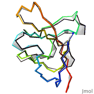
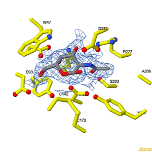
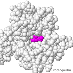

Selected Pages Art on Science Journals Education
About this image
BREAKTHROUGH in protein structure prediction!

by Eric Martz
After decades of slow progress by many groups, in 2020, AlphaFold2 proved able to accurately predict the detailed structures of two-thirds of single protein domains from their amino acid sequences. Pictured is AlphaFold2's prediction for the ORF8 protein of SARS-CoV-2 (black), compared with a subsequently published X-ray crystallographic structure (colors). ORF8 contributes to virulence in COVID-19.
>>> Visit this page >>>

About this image
Molecular Sculpture

by Eric Martz
A historical review on sculptures and physical models of macromolecules.

>>> Visit this page >>>

About this image
Interconversion of the specificities of human lysosomal enzymes associated with Fabry and Schindler diseases.

IB Tomasic, MC Metcalf, AI Guce, NE Clark, SC Garman. J. Biol. Chem. 2010 doi: 10.1074/jbc.M110.118588
The human lysosomal enzymes α-galactosidase and α-N-acetylgalactosaminidase share 46% amino acid sequence identity and have similar folds. Using a rational protein engineering approach, we interconverted the enzymatic specificity of α-GAL and α-NAGAL. The engineered α-GAL retains the antigenicity but has acquired the enzymatic specificity of α-NAGAL. Conversely, the engineered α-NAGAL retains the antigenicity but has acquired the enzymatic specificity of the α-GAL enzyme. Comparison of the crystal structures of the designed enzyme to the wild-type enzymes shows that active sites superimpose well, indicating success of the rational design. The designed enzymes might be useful as non-immunogenic alternatives in enzyme replacement therapy for treatment of lysosomal storage disorders such as Fabry disease.

>>> Visit this I3DC complement >>>

About this image
Transport of Drugs & Nutrients

Above is a transmembrane protein that takes up, into your intestinal cells, orally consumed peptide nutrients and drugs. Its lumen-face (shown above) opens and binds peptide or drug, then closes, while its cytoplasmic face (opposite end from the above) opens to release its cargo into the intestinal cell, which passes it on into the blood circulation.

>>> See more animations and explanation >>>

How to add content to Proteopedia

Video Guides

Who knows ...

List of Art on Science pages in Proteopedia

What is an Interactive 3D Complement (I3DC)?

List of I3DCs

How to get an I3DC for your paper

Teaching Strategies Using Proteopedia

Examples of Pages for Teaching

How to add content to Proteopedia

About Contact Table of Contents Structure Index Help

Proteopedia Page Contributors and Editors (what is this?)

Joel L. Sussman, Jaime Prilusky

Personal tools