Calcium uptake protein 1
From Proteopedia
(Difference between revisions)
| (12 intermediate revisions not shown.) | |||
| Line 1: | Line 1: | ||
==Structure of MICU1== | ==Structure of MICU1== | ||
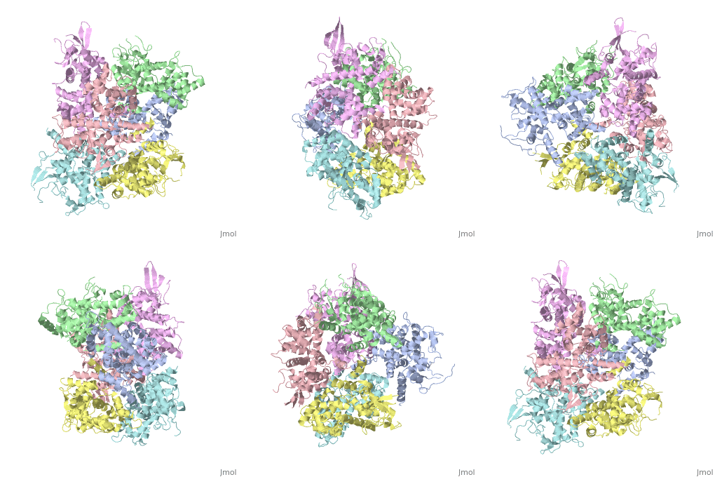
| - | <StructureSection load='4NSC' size='340' side=' | + | <StructureSection load='4NSC' size='340' side='left' caption='Human mitochondrial calcium uptake protein 1 (PDB code [[4nsc]])' scene=''> |
| - | MICU 1 (Mitochondrial Calcium Uptake 1) is a key regulator of mitochondrial calcium uniporter (MCU) required to increase calcium uptake by MCU when cytoplasmic calcium is high. | + | '''MICU 1''' ('''Mitochondrial Calcium Uptake Protein 1''') is a key regulator of mitochondrial calcium uniporter (MCU) required to increase calcium uptake by MCU when cytoplasmic calcium is high. |
It also regulates glucose-dependent insulin secretion in pancreatic beta-cells by regulating mitochondrial calcium uptake. | It also regulates glucose-dependent insulin secretion in pancreatic beta-cells by regulating mitochondrial calcium uptake. | ||
| Line 11: | Line 11: | ||
</table> | </table> | ||
| + | == History == | ||
| + | MICU1 was identified as an essential element of mitochondrial calcium uptake in 2010. It was crystallise in the presence of calcium chloride and methyl-pentanediol (MPD). Recent discover of this protein doesn’t permit to have more information about its evolutionary conservation. | ||
== Function == | == Function == | ||
| Line 20: | Line 22: | ||
== Structural highlights == | == Structural highlights == | ||
| + | MICU 1 is a ~54kDa transmembrane protein composed by an amino-terminal mitochondrial targeting sequence, a transmembrane helix and a cytosolic C-terminus which contain different important domains describe after. | ||
| + | |||
In absence of Ca2+, MICU1 is an homohexamer with a <scene name='72/723172/Secondary_structure_of_micu_1/1'>secondary structure</scene> composed by several alpha helix, beta sheets and loops. When it <scene name='72/723172/Protein_dimer_when_ca_is_bound/3'>links to this ion</scene> , it becomes a homooligomer. | In absence of Ca2+, MICU1 is an homohexamer with a <scene name='72/723172/Secondary_structure_of_micu_1/1'>secondary structure</scene> composed by several alpha helix, beta sheets and loops. When it <scene name='72/723172/Protein_dimer_when_ca_is_bound/3'>links to this ion</scene> , it becomes a homooligomer. | ||
MICU 1 is composed by differents domains : | MICU 1 is composed by differents domains : | ||
| - | :- C helix region (or Coiled-coil domain) : this domain permits different interactions with other proteins (MCU/MICU2) and is required to assemble free Ca2+ with homohexamer. | + | :- <scene name='72/723172/C-helix_region/3'>C-helix region</scene> (or Coiled-coil domain) : this domain permits different interactions with other proteins (MCU/MICU2) and is required to assemble free Ca2+ with homohexamer. |
| - | :- Polybasic region : it’s a domain which permits protein-protein interactions | + | :- <scene name='72/723172/Polybasic_region/1'>Polybasic region</scene> : it’s a domain which permits protein-protein interactions |
:- <scene name='72/723172/Ef_domains/1'>EF-hand domains</scene> There are 2 of them. They are the most important domains in MICU1 protein because they contain the Ca2+ binding region and detect the concentration of calcium in the cell for activation of signalling. An EF-hand domain is a calcium sensor. An EF-hand domain is a helix loop helix structural domain that means that it is 2 alpha helices linked by a short loop region. Ca2+ ions are coordinated in this space thanks to ligands within the loop : in EF-1 residues concerning are Asp231, Asn233, Asp235, Glu237 and Glu242 and in EF-2 they are Asp421, Asp423, Asn425, Glu427 and Glu432. When Ca2+ binds itself to this domain, the protein changes its conformation to expose a domain that can interact with other proteins. | :- <scene name='72/723172/Ef_domains/1'>EF-hand domains</scene> There are 2 of them. They are the most important domains in MICU1 protein because they contain the Ca2+ binding region and detect the concentration of calcium in the cell for activation of signalling. An EF-hand domain is a calcium sensor. An EF-hand domain is a helix loop helix structural domain that means that it is 2 alpha helices linked by a short loop region. Ca2+ ions are coordinated in this space thanks to ligands within the loop : in EF-1 residues concerning are Asp231, Asn233, Asp235, Glu237 and Glu242 and in EF-2 they are Asp421, Asp423, Asn425, Glu427 and Glu432. When Ca2+ binds itself to this domain, the protein changes its conformation to expose a domain that can interact with other proteins. | ||
== Disease == | == Disease == | ||
If MICU1 gene is modified, Ca2+ can be load in higher concentration in the mitochondry resulting Myopathy with extrapyramidal signs (MPXPS). This is an autosomal recessive disorder characterized by early-onset proximal muscle weakness with a static course and moderately to grossly elevated serum creatine kinase levels accompanied by learning difficulties. Most patients develop subtle extrapyramidal motor signs that progress to a debilitating disorder of involuntary movement with variable features, including chorea, tremor, dystonic posturing and orofacial dyskinesia. Additional variable features include ataxia, microcephaly, ophthalmoplegia, ptosis, optic atrophy and axonal peripheral neuropathy. | If MICU1 gene is modified, Ca2+ can be load in higher concentration in the mitochondry resulting Myopathy with extrapyramidal signs (MPXPS). This is an autosomal recessive disorder characterized by early-onset proximal muscle weakness with a static course and moderately to grossly elevated serum creatine kinase levels accompanied by learning difficulties. Most patients develop subtle extrapyramidal motor signs that progress to a debilitating disorder of involuntary movement with variable features, including chorea, tremor, dystonic posturing and orofacial dyskinesia. Additional variable features include ataxia, microcephaly, ophthalmoplegia, ptosis, optic atrophy and axonal peripheral neuropathy. | ||
| + | |||
| + | </StructureSection> | ||
| + | |||
| + | == 3D Structures of calcium uptake protein 1 == | ||
| + | |||
| + | [[Calcium uptake protein 3D structures]] | ||
== See Also == | == See Also == | ||
Current revision
Contents |
Structure of MICU1
| |||||||||||
3D Structures of calcium uptake protein 1
Calcium uptake protein 3D structures
See Also
References
- https://www.researchgate.net/publication/260152737_Structural_and_mechanistic_insights_into_MICU1_regulation_of_mitochondrial_calcium_uptake
- http://ghr.nlm.nih.gov/gene/MICU1
- http://www.rcsb.org/pdb/home/home.do
- http://www.uniprot.org/uniprot/Q9BPX6

