Gyrase
From Proteopedia
(Difference between revisions)
| (38 intermediate revisions not shown.) | |||
| Line 1: | Line 1: | ||
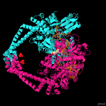
| - | + | <StructureSection load='' size='350' side='right' caption='Gyrase subunit A N-terminal (cyan) and subunit B C-terminal (magenta) complex with DNA, inhibitor, sulfate and Mn+2 ion (purple) (PDB entry [[4plb]])' scene='41/410321/Cv/1'> | |
| + | ==Function== | ||
| - | + | '''Gyrase (Gyr)''' is a type of topoisomerase II in prokaryotes which unwinds double stranded DNA. The DNA Gyr cutting allows the formation of a negative DNA supercoil which enables replication of DNA<ref>PMID:1657531</ref> Gyr consists of 2 subunits: GyrA and GyrB. '''Reverse gyrase''' (Top-RG) is a type of topoisomerase I which catalyses the formation of positive DNA supercoil. <ref>PMID:15673717</ref> See also [[Isomerases]]. | |
| - | + | ==Relevance== | |
| - | + | GyrA inhibitor [[Ciprofloxacin]] is used as antibiotic drug. Fluoroquinolones are Gyr inhibitors used in treatment of multi drug-resistant tuberculosis<ref>PMID:15047530</ref> | |
| + | |||
| + | == Structural highlights == | ||
| + | <scene name='41/410321/Cv/5'>Gyrase subunit A N-terminal and subunit B C-terminal complex with DNA and inhibitor</scene>. A <scene name='41/410321/Cv/6'>potential drug interacts with both DNA strands and in the hydrophobic pocket between subunits A and B</scene><ref>PMID:24900889</ref>. Water molecules shown as red spheres. | ||
==3D Structure of Gyrase== | ==3D Structure of Gyrase== | ||
| + | [[Gyrase 3D Structures]] | ||
| - | + | </StructureSection> | |
| - | + | ||
| - | + | ||
| - | + | ||
| - | + | ||
| - | + | ||
| - | + | ||
| - | + | ||
| - | + | ||
| - | + | ||
| - | + | ||
| - | + | ||
| - | + | ||
| - | + | ||
| - | + | ||
| - | + | ||
| - | + | ||
| - | + | ||
| - | + | ||
| - | + | ||
| - | + | ||
| - | + | ||
| - | + | ||
| - | + | ||
| - | + | ||
| - | + | ||
| - | + | ||
| - | + | ||
| - | + | ||
| - | + | ||
| - | + | ||
| - | + | ||
| - | + | ||
| - | + | ||
==Additional Resources== | ==Additional Resources== | ||
Current revision
| |||||||||||
Additional Resources
For additional information, see: Bacterial Infections
References
- ↑ Reece RJ, Maxwell A. DNA gyrase: structure and function. Crit Rev Biochem Mol Biol. 1991;26(3-4):335-75. PMID:1657531 doi:http://dx.doi.org/10.3109/10409239109114072
- ↑ Napoli A, Valenti A, Salerno V, Nadal M, Garnier F, Rossi M, Ciaramella M. Functional interaction of reverse gyrase with single-strand binding protein of the archaeon Sulfolobus. Nucleic Acids Res. 2005 Jan 26;33(2):564-76. Print 2005. PMID:15673717 doi:http://dx.doi.org/10.1093/nar/gki202
- ↑ Aubry A, Pan XS, Fisher LM, Jarlier V, Cambau E. Mycobacterium tuberculosis DNA gyrase: interaction with quinolones and correlation with antimycobacterial drug activity. Antimicrob Agents Chemother. 2004 Apr;48(4):1281-8. PMID:15047530
- ↑ Singh SB, Kaelin DE, Wu J, Miesel L, Tan CM, Meinke PT, Olsen D, Lagrutta A, Bradley P, Lu J, Patel S, Rickert KW, Smith RF, Soisson S, Wei C, Fukuda H, Kishii R, Takei M, Fukuda Y. Oxabicyclooctane-linked novel bacterial topoisomerase inhibitors as broad spectrum antibacterial agents. ACS Med Chem Lett. 2014 Mar 12;5(5):609-14. doi: 10.1021/ml500069w. eCollection, 2014 May 8. PMID:24900889 doi:http://dx.doi.org/10.1021/ml500069w
Proteopedia Page Contributors and Editors (what is this?)
Michal Harel, Alexander Berchansky, David Canner, Joel L. Sussman
