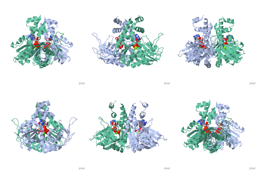
Plasmid segregation protein ParM
From Proteopedia
(Difference between revisions)
| (2 intermediate revisions not shown.) | |||
| Line 4: | Line 4: | ||
== Structural highlights == | == Structural highlights == | ||
| - | ParM polymerizes into double helical protofilaments with repeats similar to F-actin. <scene name='71/715423/Cv/ | + | ParM polymerizes into double helical protofilaments with repeats similar to F-actin. <scene name='71/715423/Cv/4'>AMPPNP binding site</scene>. |
| - | *<scene name='71/715423/Cv/ | + | *<scene name='71/715423/Cv/5'>Mg coordination site</scene>. Water molecules are shown as red spheres. |
| Line 25: | Line 25: | ||
== References == | == References == | ||
<references/> | <references/> | ||
| + | [[Category:Topic Page]] | ||
Current revision
| |||||||||||
3D structures of plasmid segregation protein ParM
Updated on 07-August-2019
1mwk – ParM – Escherichias coli
1mwm – ParM + ADP
2zgy – ParM + GDP
2zgz – ParM + GMPPNP
4a61 – ParM + AMPPNP
5aey – ParM + AMPPNP - CryoEM
4a62 – ParM + ParR C terminus
2qu4, 2ghc, 3iku, 3iky, 4a6j, 5ai7 – ParM – CryoEM
2zhc – ParM + ADP - CryoEM
References
- ↑ Salje J, Gayathri P, Lowe J. The ParMRC system: molecular mechanisms of plasmid segregation by actin-like filaments. Nat Rev Microbiol. 2010 Oct;8(10):683-92. doi: 10.1038/nrmicro2425. PMID:20844556 doi:http://dx.doi.org/10.1038/nrmicro2425

