Main Page

From Proteopedia

(Difference between revisions)
Jump to: navigation, search
(Undo revision 3114449 by Deja Sanders (Talk))
Line 1: Line 1:
-
<table id="Tetrahydroprotoberberine N-methyltransferase" style="width:100%;border:2px solid #ddd;border-collapse: collapse;table-layout: fixed; ">
+
<table id="tableColumnsMainPage" style="width:100%;border:2px solid #ddd;border-collapse: collapse;table-layout: fixed; ">
<tr><td colspan='3' style="background:#F5F5FC;border:1px solid #ddd;">
<div style="top:+0.2em; font-size:1.2em; padding:5px 5px 5px 10px; float:right;">'''''ISSN 2310-6301'''''</div>

Revision as of 05:43, 26 November 2019

ISSN 2310-6301

As life is more than 2D, Proteopedia helps to bridge the gap between 3D structure & function of biomacromolecules

Proteopedia presents this information in a user-friendly way as a collaborative & free 3D-encyclopedia of proteins & other biomolecules.


Selected Research Pages In Journals Education
About this image
Coronavirus Spike Protein Priming

by Eric Martz
Coronavirus SARS-CoV-2 (responsible for COVID-19) has a spike protein on its surface, which enables it to infect host cells. Initially, proteases in the lungs clip the homo-trimeric spike protein at a unique sequence. This primes it, causing it to extend its receptor binding surface (shown in the above animation), optimizing binding to the host cell's ACE2 receptor (not shown). Next, spike protein initiates fusion of the virus and host cell membranes (not shown), enabling the virus RNA to enter the cell and initiate production of new virions. Knowledge of spike protein's molecular structure and function is crucial to developing effective therapies and vaccines.
>>> Visit this page >>>

About this image
Geobacter nanowire structure surprise.

F Wang, Y Gu, JP O'Brien, SM Yi, SE Yalcin, V Srikanth, C Shen, D Vu, NL Ing, AI Hochbaum, EH Egelman, NS Malvankar. Cell 2019 doi: 10.1016/j.cell.2019.03.029
Bacteria living in anaerobic environments (no oxygen) need alternative electron acceptors in order to get energy from their food. An acceptor abundant in the earth's crust is red iron oxide ("rust"), which gets reduced to black iron oxide (magnetite). Many bacteria, such as Geobacter, get their metabolic energy by transferring electrons to acceptors that are multiple cell diameters distant, using protein nanowires. These were long thought to be pili. But when the structure of the nanowires was solved in 2019, to everyone's surprise, they turned out to be unprecedented linear polymers of multi-heme cytochromes. The hemes form an electrically conductive chain in the cores of these nanowires.

>>> Visit I3DC Interactive Visualizations >>>

About this image
Tutorial: Ramachandran Plot Inspection

by Angel Herráez
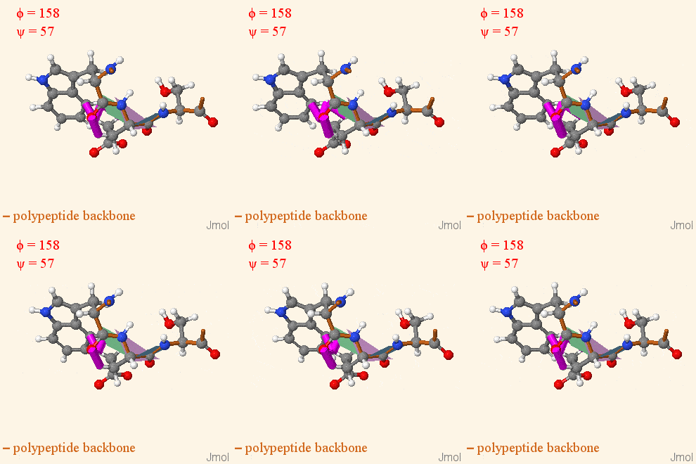
Side-by-side display of dihedral angles in a 3D model of a tripeptide and its Ramachandran plot. Users can interact with any of them and the other will change accordingly. Includes animated rotations with display of clashes.

>>> Visit this page >>>

How to add content to Proteopedia

Video Guides

Who knows ...

About Interactive 3D Complements - I3DCs

List of I3DCs

How to get an I3DC for your paper

Teaching strategies using Proteopedia

Examples of pages for teaching

How to add content to Proteopedia

About Contact Hot News Table of Contents Structure Index Help

Proteopedia Page Contributors and Editors (what is this?)

Joel L. Sussman, Jaime Prilusky

Personal tools