1cfc
From Proteopedia
| Line 1: | Line 1: | ||
[[Image:1cfc.gif|left|200px]] | [[Image:1cfc.gif|left|200px]] | ||
| - | + | <!-- | |
| - | + | The line below this paragraph, containing "STRUCTURE_1cfc", creates the "Structure Box" on the page. | |
| - | + | You may change the PDB parameter (which sets the PDB file loaded into the applet) | |
| - | + | or the SCENE parameter (which sets the initial scene displayed when the page is loaded), | |
| - | | | + | or leave the SCENE parameter empty for the default display. |
| - | | | + | --> |
| - | + | {{STRUCTURE_1cfc| PDB=1cfc | SCENE= }} | |
| - | + | ||
| - | + | ||
| - | }} | + | |
'''CALCIUM-FREE CALMODULIN''' | '''CALCIUM-FREE CALMODULIN''' | ||
| Line 31: | Line 28: | ||
[[Category: Ren, H.]] | [[Category: Ren, H.]] | ||
[[Category: Tjandra, N.]] | [[Category: Tjandra, N.]] | ||
| - | [[Category: | + | [[Category: Calcium-binding protein]] |
| - | + | ''Page seeded by [http://oca.weizmann.ac.il/oca OCA ] on Fri May 2 12:40:28 2008'' | |
| - | ''Page seeded by [http://oca.weizmann.ac.il/oca OCA ] on | + | |
Revision as of 09:40, 2 May 2008
| |||||||||
| 1cfc, 25 NMR models () | |||||||||
|---|---|---|---|---|---|---|---|---|---|
| Related: | 1cfd | ||||||||
| |||||||||
| |||||||||
| |||||||||
| Resources: | FirstGlance, OCA, RCSB, PDBsum | ||||||||
| Coordinates: | save as pdb, mmCIF, xml | ||||||||
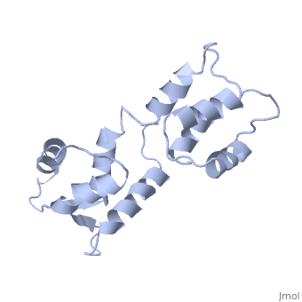

CALCIUM-FREE CALMODULIN
Overview
The three-dimensional structure of calmodulin in the absence of Ca2+ has been determined by three- and four-dimensional heteronuclear NMR experiments, including ROE, isotope-filtering combined with reverse labelling, and measurement of more than 700 three-bond J-couplings. In analogy with the Ca(2+)-ligated state of this protein, it consists of two small globular domains separated by a flexible linker, with no stable, direct contacts between the two domains. In the absence of Ca2+, the four helices in each of the two globular domains form a highly twisted bundle, capped by a short anti-parallel beta-sheet. This arrangement is qualitatively similar to that observed in the crystal structure of the Ca(2+)-free N-terminal domain of troponin C.
About this Structure
1CFC is a Single protein structure of sequence from Xenopus laevis. Full crystallographic information is available from OCA.
Reference
Solution structure of calcium-free calmodulin., Kuboniwa H, Tjandra N, Grzesiek S, Ren H, Klee CB, Bax A, Nat Struct Biol. 1995 Sep;2(9):768-76. PMID:7552748 Page seeded by OCA on Fri May 2 12:40:28 2008
