1e2h
From Proteopedia
| Line 1: | Line 1: | ||
[[Image:1e2h.gif|left|200px]] | [[Image:1e2h.gif|left|200px]] | ||
| - | + | <!-- | |
| - | + | The line below this paragraph, containing "STRUCTURE_1e2h", creates the "Structure Box" on the page. | |
| - | + | You may change the PDB parameter (which sets the PDB file loaded into the applet) | |
| - | + | or the SCENE parameter (which sets the initial scene displayed when the page is loaded), | |
| - | + | or leave the SCENE parameter empty for the default display. | |
| - | | | + | --> |
| - | | | + | {{STRUCTURE_1e2h| PDB=1e2h | SCENE= }} |
| - | + | ||
| - | + | ||
| - | }} | + | |
'''THE NUCLEOSIDE BINDING SITE OF HERPES SIMPLEX TYPE 1 THYMIDINE KINASE ANALYZED BY X-RAY CRYSTALLOGRAPHY''' | '''THE NUCLEOSIDE BINDING SITE OF HERPES SIMPLEX TYPE 1 THYMIDINE KINASE ANALYZED BY X-RAY CRYSTALLOGRAPHY''' | ||
| Line 29: | Line 26: | ||
[[Category: Schulz, G E.]] | [[Category: Schulz, G E.]] | ||
[[Category: Vogt, J.]] | [[Category: Vogt, J.]] | ||
| - | [[Category: | + | [[Category: Adenine analog]] |
| - | [[Category: | + | [[Category: Enzyme-prodrug gene therapy]] |
| - | [[Category: | + | [[Category: Nucleoside-binding]] |
| - | [[Category: | + | [[Category: Thymidine kinase]] |
| - | [[Category: | + | [[Category: X-ray crystallography]] |
| - | + | ''Page seeded by [http://oca.weizmann.ac.il/oca OCA ] on Fri May 2 14:34:37 2008'' | |
| - | ''Page seeded by [http://oca.weizmann.ac.il/oca OCA ] on | + | |
Revision as of 11:34, 2 May 2008
| |||||||||
| 1e2h, resolution 1.90Å () | |||||||||
|---|---|---|---|---|---|---|---|---|---|
| Ligands: | |||||||||
| Activity: | Thymidine kinase, with EC number 2.7.1.21 | ||||||||
| Related: | 1kim, 1vtk, 2vtk, 3vtk, 1ki2, 1ki4, 1ki5, 1ki6, 1ki7, 1ki8, 1e2i, 1e2j, 1e2k, 1e2l, 1e2m, 1e2n, 1e2p | ||||||||
| |||||||||
| |||||||||
| |||||||||
| Resources: | FirstGlance, OCA, RCSB, PDBsum | ||||||||
| Coordinates: | save as pdb, mmCIF, xml | ||||||||

THE NUCLEOSIDE BINDING SITE OF HERPES SIMPLEX TYPE 1 THYMIDINE KINASE ANALYZED BY X-RAY CRYSTALLOGRAPHY
Overview
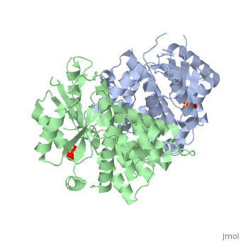
The crystal structures of the full-length Herpes simplex virus type 1 thymidine kinase in its unligated form and in a complex with an adenine analogue have been determined at 1.9 A resolution. The unligated enzyme contains four water molecules in the thymidine pocket and reveals a small induced fit on substrate binding. The structure of the ligated enzyme shows for the first time a bound adenine analogue after numerous complexes with thymine and guanine analogues have been reported. The adenine analogue constitutes a new lead compound for enzyme-prodrug gene therapy. In addition, the structure of mutant Q125N modifying the binding site of the natural substrate thymidine in complex with this substrate has been established at 2.5 A resolution. It reveals that neither the binding mode of thymidine nor the polypeptide backbone conformation is altered, except that the two major hydrogen bonds to thymidine are replaced by a single water-mediated hydrogen bond, which improves the relative acceptance of the prodrugs aciclovir and ganciclovir compared with the natural substrate. Accordingly, the mutant structure represents a first step toward improving the virus-directed enzyme-prodrug gene therapy by enzyme engineering.
About this Structure
1E2H is a Single protein structure of sequence from Human herpesvirus 1. Full crystallographic information is available from OCA.
Reference
Nucleoside binding site of herpes simplex type 1 thymidine kinase analyzed by X-ray crystallography., Vogt J, Perozzo R, Pautsch A, Prota A, Schelling P, Pilger B, Folkers G, Scapozza L, Schulz GE, Proteins. 2000 Dec 1;41(4):545-53. PMID:11056041 Page seeded by OCA on Fri May 2 14:34:37 2008
