Journal:IUCrJ:S205225252000072X
From Proteopedia
(Difference between revisions)

| (3 intermediate revisions not shown.) | |||
| Line 1: | Line 1: | ||
<StructureSection load='' size='370' side='right' scene='83/834718/Cv/5' caption=''> | <StructureSection load='' size='370' side='right' scene='83/834718/Cv/5' caption=''> | ||
===Comparing serial X-ray crystallography and microcrystal electron diffraction (MicroED) as methods for routine structure determination from small macromolecular crystals=== | ===Comparing serial X-ray crystallography and microcrystal electron diffraction (MicroED) as methods for routine structure determination from small macromolecular crystals=== | ||
| - | <big>Alexander M. Wolff, Iris D. Young, Raymond G. Sierra, Aaron S. Brewster, Michael W. Martynowycz, Eriko Nango, Michihiro Sugahara, Takanori Nakane, Kazutaka Ito, Andrew Aquila, Asmit Bhowmik, Justin T. Biel, Sergio Carbajo, Aina E. Cohen | + | <big>Alexander M. Wolff, Iris D. Young, Raymond G. Sierra, Aaron S. Brewster, Michael W. Martynowycz, Eriko Nango, Michihiro Sugahara, Takanori Nakane, Kazutaka Ito, Andrew Aquila, Asmit Bhowmik, Justin T. Biel, Sergio Carbajo, Aina E. Cohen, Saul Cortez, Ana Gonzalez, Tomoya Hino, Dohyun Im, Jake D. Koralek, Minoru Kubo, Tomas S. Lazarou, Takashi Nomura, Shigeki Owada, Avi J. Samelson, Tomoyuki Tanaka, Rie Tanaka, Erin M. Thompson, Henry van den Bedem, Rahel A. Woldeyes, Fumiaki Yumoto, Wei Zhao, Kensuke Tono, Sébastien Boutet, So Iwata, Tamir Gonen, Nicholas K. Sauter, James S. Fraser, Michael C. Thompson</big> <ref>doi 10.1107/S205225252000072X</ref> |
<hr/> | <hr/> | ||
<b>Molecular Tour</b><br> | <b>Molecular Tour</b><br> | ||
| Line 13: | Line 13: | ||
{{Clear}} | {{Clear}} | ||
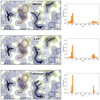
Comparison of the 2mFoFc map and the refined multi-conformer model produced from each serial XFEL experiment. Maps were visualized at multiple contour levels to show evidence for alternative conformations (see static image above). Following multi-conformer refinement, ensembles were generated from each model using phenix.ensemble_refine. In the right panel, a histogram of the Chi1 angles for residue 113 is plotted for the ensemble. Multi-conformer models plus maps, and the distribution of Chi1 angles across the ensemble models are similar for all three XFEL data sets: | Comparison of the 2mFoFc map and the refined multi-conformer model produced from each serial XFEL experiment. Maps were visualized at multiple contour levels to show evidence for alternative conformations (see static image above). Following multi-conformer refinement, ensembles were generated from each model using phenix.ensemble_refine. In the right panel, a histogram of the Chi1 angles for residue 113 is plotted for the ensemble. Multi-conformer models plus maps, and the distribution of Chi1 angles across the ensemble models are similar for all three XFEL data sets: | ||
| - | *<scene name='83/834718/Cv/2'>XFEL MESH</scene>. | + | *<scene name='83/834718/Cv/2'>XFEL MESH</scene> ([[6u5c]]). |
| - | *<scene name='83/834718/Cv/3'>XFEL LCP</scene>. | + | *<scene name='83/834718/Cv/3'>XFEL LCP</scene> ([[6u5d]]). |
| - | *<scene name='83/834718/Cv/4'>XFEL Cellulose</scene> | + | *<scene name='83/834718/Cv/4'>XFEL Cellulose</scene> ([[6u5e]]). |
[[Image:fig4_alt_revised.001.png|left|400px|thumb]] | [[Image:fig4_alt_revised.001.png|left|400px|thumb]] | ||
{{Clear}} | {{Clear}} | ||
Visualization of the 2mFoFc map and the refined model measured from a FIB-milled crystal using MicroED (see static image above). The conformation of residues coupled to the catalytic site resembles structures previously solved under cryogenic conditions using X-ray crystallography (PDB ID [[3k0m]]). For some regions of the structure, the cryogenic X-ray and MicroED structures are indistinguishable. A previously published multi-conformer model produced from data acquired at room temperature is provided for comparison (PDB ID [[3k0n]]). Following refinement, ensembles were generated using phenix.ensemble_refine. In the right panel, a histogram of the Chi1 angles for residue 113 is plotted for the ensemble. All members of the ensemble adopted the same rotameric position as previous cryogenic structures. | Visualization of the 2mFoFc map and the refined model measured from a FIB-milled crystal using MicroED (see static image above). The conformation of residues coupled to the catalytic site resembles structures previously solved under cryogenic conditions using X-ray crystallography (PDB ID [[3k0m]]). For some regions of the structure, the cryogenic X-ray and MicroED structures are indistinguishable. A previously published multi-conformer model produced from data acquired at room temperature is provided for comparison (PDB ID [[3k0n]]). Following refinement, ensembles were generated using phenix.ensemble_refine. In the right panel, a histogram of the Chi1 angles for residue 113 is plotted for the ensemble. All members of the ensemble adopted the same rotameric position as previous cryogenic structures. | ||
| - | *<scene name='83/834718/Cv/ | + | *<scene name='83/834718/Cv/7'>The refined model measured from a FIB-milled crystal using MicroED</scene> ([[6u5g]], colored in magenta). [[3k0n]] (room temp X-ray) is in orange and [[3k0m]] (cryo X-ray) is in deepskyblue. |
| + | |||
<b>References</b><br> | <b>References</b><br> | ||
<references/> | <references/> | ||
</StructureSection> | </StructureSection> | ||
__NOEDITSECTION__ | __NOEDITSECTION__ | ||
Current revision
| |||||||||||
This page complements a publication in scientific journals and is one of the Proteopedia's Interactive 3D Complement pages. For aditional details please see I3DC.


