This old version of Proteopedia is provided for student assignments while the new version is undergoing repairs. Content and edits done in this old version of Proteopedia after March 1, 2026 will eventually be lost when it is retired in about June of 2026.
Apply for new accounts at the new Proteopedia. Your logins will work in both the old and new versions.
3ku2
From Proteopedia
(Difference between revisions)
| (8 intermediate revisions not shown.) | |||
| Line 1: | Line 1: | ||
| - | {{Seed}} | ||
| - | [[Image:3ku2.png|left|200px]] | ||
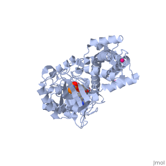
| - | < | + | ==Crystal Structure of inactivated form of CDPK1 from toxoplasma gondii, TGME49.101440== |
| - | + | <StructureSection load='3ku2' size='340' side='right'caption='[[3ku2]], [[Resolution|resolution]] 2.30Å' scene=''> | |
| - | You may | + | == Structural highlights == |
| - | + | <table><tr><td colspan='2'>[[3ku2]] is a 1 chain structure with sequence from [https://en.wikipedia.org/wiki/Toxoplasma_gondii Toxoplasma gondii]. Full crystallographic information is available from [http://oca.weizmann.ac.il/oca-bin/ocashort?id=3KU2 OCA]. For a <b>guided tour on the structure components</b> use [https://proteopedia.org/fgij/fg.htm?mol=3KU2 FirstGlance]. <br> | |
| - | + | </td></tr><tr id='method'><td class="sblockLbl"><b>[[Empirical_models|Method:]]</b></td><td class="sblockDat" id="methodDat">X-ray diffraction, [[Resolution|Resolution]] 2.3Å</td></tr> | |
| - | - | + | <tr id='ligand'><td class="sblockLbl"><b>[[Ligand|Ligands:]]</b></td><td class="sblockDat" id="ligandDat"><scene name='pdbligand=ANP:PHOSPHOAMINOPHOSPHONIC+ACID-ADENYLATE+ESTER'>ANP</scene>, <scene name='pdbligand=UNX:UNKNOWN+ATOM+OR+ION'>UNX</scene></td></tr> |
| - | + | <tr id='resources'><td class="sblockLbl"><b>Resources:</b></td><td class="sblockDat"><span class='plainlinks'>[https://proteopedia.org/fgij/fg.htm?mol=3ku2 FirstGlance], [http://oca.weizmann.ac.il/oca-bin/ocaids?id=3ku2 OCA], [https://pdbe.org/3ku2 PDBe], [https://www.rcsb.org/pdb/explore.do?structureId=3ku2 RCSB], [https://www.ebi.ac.uk/pdbsum/3ku2 PDBsum], [https://prosat.h-its.org/prosat/prosatexe?pdbcode=3ku2 ProSAT]</span></td></tr> | |
| + | </table> | ||
| + | == Function == | ||
| + | [https://www.uniprot.org/uniprot/Q9BJF5_TOXGO Q9BJF5_TOXGO] | ||
| + | == Evolutionary Conservation == | ||
| + | [[Image:Consurf_key_small.gif|200px|right]] | ||
| + | Check<jmol> | ||
| + | <jmolCheckbox> | ||
| + | <scriptWhenChecked>; select protein; define ~consurf_to_do selected; consurf_initial_scene = true; script "/wiki/ConSurf/ku/3ku2_consurf.spt"</scriptWhenChecked> | ||
| + | <scriptWhenUnchecked>script /wiki/extensions/Proteopedia/spt/initialview01.spt</scriptWhenUnchecked> | ||
| + | <text>to colour the structure by Evolutionary Conservation</text> | ||
| + | </jmolCheckbox> | ||
| + | </jmol>, as determined by [http://consurfdb.tau.ac.il/ ConSurfDB]. You may read the [[Conservation%2C_Evolutionary|explanation]] of the method and the full data available from [http://bental.tau.ac.il/new_ConSurfDB/main_output.php?pdb_ID=3ku2 ConSurf]. | ||
| + | <div style="clear:both"></div> | ||
| + | <div style="background-color:#fffaf0;"> | ||
| + | == Publication Abstract from PubMed == | ||
| + | Calcium-dependent protein kinases (CDPKs) have pivotal roles in the calcium-signaling pathway in plants, ciliates and apicomplexan parasites and comprise a calmodulin-dependent kinase (CaMK)-like kinase domain regulated by a calcium-binding domain in the C terminus. To understand this intramolecular mechanism of activation, we solved the structures of the autoinhibited (apo) and activated (calcium-bound) conformations of CDPKs from the apicomplexan parasites Toxoplasma gondii and Cryptosporidium parvum. In the apo form, the C-terminal CDPK activation domain (CAD) resembles a calmodulin protein with an unexpected long helix in the N terminus that inhibits the kinase domain in the same manner as CaMKII. Calcium binding triggers the reorganization of the CAD into a highly intricate fold, leading to its relocation around the base of the kinase domain to a site remote from the substrate binding site. This large conformational change constitutes a distinct mechanism in calcium signal-transduction pathways. | ||
| - | + | Structures of apicomplexan calcium-dependent protein kinases reveal mechanism of activation by calcium.,Wernimont AK, Artz JD, Finerty P Jr, Lin YH, Amani M, Allali-Hassani A, Senisterra G, Vedadi M, Tempel W, Mackenzie F, Chau I, Lourido S, Sibley LD, Hui R Nat Struct Mol Biol. 2010 May;17(5):596-601. Epub 2010 May 2. PMID:20436473<ref>PMID:20436473</ref> | |
| + | From MEDLINE®/PubMed®, a database of the U.S. National Library of Medicine.<br> | ||
| + | </div> | ||
| + | <div class="pdbe-citations 3ku2" style="background-color:#fffaf0;"></div> | ||
| - | == | + | ==See Also== |
| - | + | *[[Calcium-dependent protein kinase|Calcium-dependent protein kinase]] | |
| + | == References == | ||
| + | <references/> | ||
| + | __TOC__ | ||
| + | </StructureSection> | ||
| + | [[Category: Large Structures]] | ||
[[Category: Toxoplasma gondii]] | [[Category: Toxoplasma gondii]] | ||
| - | [[Category: Arrowsmith | + | [[Category: Arrowsmith CH]] |
| - | [[Category: Artz | + | [[Category: Artz JD]] |
| - | [[Category: Bochkarev | + | [[Category: Bochkarev A]] |
| - | [[Category: Bountra | + | [[Category: Bountra C]] |
| - | [[Category: Edwards | + | [[Category: Edwards AM]] |
| - | [[Category: Finnerty | + | [[Category: Finnerty P]] |
| - | [[Category: Hassani | + | [[Category: Hassani AA]] |
| - | [[Category: He | + | [[Category: He H]] |
| - | [[Category: Hui | + | [[Category: Hui R]] |
| - | [[Category: | + | [[Category: Lin YH]] |
| - | [[Category: | + | [[Category: Lourido S]] |
| - | [[Category: | + | [[Category: Mackenzie F]] |
| - | [[Category: Sibley | + | [[Category: Sibley DL]] |
| - | [[Category: Sinestera | + | [[Category: Sinestera G]] |
| - | [[Category: Vedadi | + | [[Category: Vedadi M]] |
| - | [[Category: Wasney | + | [[Category: Wasney G]] |
| - | [[Category: Weigelt | + | [[Category: Weigelt J]] |
| - | [[Category: Wernimont | + | [[Category: Wernimont AK]] |
| - | [[Category: Xiao | + | [[Category: Xiao T]] |
| - | + | ||
| - | + | ||
| - | + | ||
| - | + | ||
| - | + | ||
| - | + | ||
| - | + | ||
| - | + | ||
| - | + | ||
| - | + | ||
| - | + | ||
| - | + | ||
| - | + | ||
Current revision
Crystal Structure of inactivated form of CDPK1 from toxoplasma gondii, TGME49.101440
| |||||||||||

Categories: Large Structures | Toxoplasma gondii | Arrowsmith CH | Artz JD | Bochkarev A | Bountra C | Edwards AM | Finnerty P | Hassani AA | He H | Hui R | Lin YH | Lourido S | Mackenzie F | Sibley DL | Sinestera G | Vedadi M | Wasney G | Weigelt J | Wernimont AK | Xiao T
