This old version of Proteopedia is provided for student assignments while the new version is undergoing repairs. Content and edits done in this old version of Proteopedia after March 1, 2026 will eventually be lost when it is retired in about June of 2026.
Apply for new accounts at the new Proteopedia. Your logins will work in both the old and new versions.
1vgj
From Proteopedia
(Difference between revisions)
| (10 intermediate revisions not shown.) | |||
| Line 1: | Line 1: | ||
| - | [[Image:1vgj.gif|left|200px]] | ||
| - | < | + | ==Crystal structure of 2'-5' RNA ligase from Pyrococcus horikoshii== |
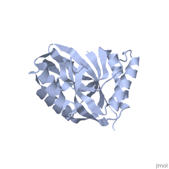
| - | + | <StructureSection load='1vgj' size='340' side='right'caption='[[1vgj]], [[Resolution|resolution]] 1.94Å' scene=''> | |
| - | You may | + | == Structural highlights == |
| - | + | <table><tr><td colspan='2'>[[1vgj]] is a 1 chain structure with sequence from [https://en.wikipedia.org/wiki/Pyrococcus_horikoshii Pyrococcus horikoshii]. Full crystallographic information is available from [http://oca.weizmann.ac.il/oca-bin/ocashort?id=1VGJ OCA]. For a <b>guided tour on the structure components</b> use [https://proteopedia.org/fgij/fg.htm?mol=1VGJ FirstGlance]. <br> | |
| - | + | </td></tr><tr id='method'><td class="sblockLbl"><b>[[Empirical_models|Method:]]</b></td><td class="sblockDat" id="methodDat">X-ray diffraction, [[Resolution|Resolution]] 1.94Å</td></tr> | |
| - | -- | + | <tr id='ligand'><td class="sblockLbl"><b>[[Ligand|Ligands:]]</b></td><td class="sblockDat" id="ligandDat"><scene name='pdbligand=MSE:SELENOMETHIONINE'>MSE</scene></td></tr> |
| - | + | <tr id='resources'><td class="sblockLbl"><b>Resources:</b></td><td class="sblockDat"><span class='plainlinks'>[https://proteopedia.org/fgij/fg.htm?mol=1vgj FirstGlance], [http://oca.weizmann.ac.il/oca-bin/ocaids?id=1vgj OCA], [https://pdbe.org/1vgj PDBe], [https://www.rcsb.org/pdb/explore.do?structureId=1vgj RCSB], [https://www.ebi.ac.uk/pdbsum/1vgj PDBsum], [https://prosat.h-its.org/prosat/prosatexe?pdbcode=1vgj ProSAT]</span></td></tr> | |
| + | </table> | ||
| + | == Function == | ||
| + | [https://www.uniprot.org/uniprot/THPR_PYRHO THPR_PYRHO] Hydrolyzes RNA 2',3'-cyclic phosphodiester to an RNA 2'-phosphomonoester.[HAMAP-Rule:MF_01940] | ||
| + | == Evolutionary Conservation == | ||
| + | [[Image:Consurf_key_small.gif|200px|right]] | ||
| + | Check<jmol> | ||
| + | <jmolCheckbox> | ||
| + | <scriptWhenChecked>; select protein; define ~consurf_to_do selected; consurf_initial_scene = true; script "/wiki/ConSurf/vg/1vgj_consurf.spt"</scriptWhenChecked> | ||
| + | <scriptWhenUnchecked>script /wiki/extensions/Proteopedia/spt/initialview01.spt</scriptWhenUnchecked> | ||
| + | <text>to colour the structure by Evolutionary Conservation</text> | ||
| + | </jmolCheckbox> | ||
| + | </jmol>, as determined by [http://consurfdb.tau.ac.il/ ConSurfDB]. You may read the [[Conservation%2C_Evolutionary|explanation]] of the method and the full data available from [http://bental.tau.ac.il/new_ConSurfDB/main_output.php?pdb_ID=1vgj ConSurf]. | ||
| + | <div style="clear:both"></div> | ||
| + | <div style="background-color:#fffaf0;"> | ||
| + | == Publication Abstract from PubMed == | ||
| + | Bacterial and archaeal 2'-5' RNA ligases, members of the 2H phosphoesterase superfamily, catalyze the linkage of the 5' and 3' exons via a 2'-5'-phosphodiester bond during tRNA-precursor splicing. The crystal structure of the 2'-5' RNA ligase PH0099 from Pyrococcus horikoshii OT3 was solved at 1.94 A resolution (PDB code 1vgj). The molecule has a bilobal alpha+beta arrangement with two antiparallel beta-sheets constituting a V-shaped active-site cleft, as found in other members of the 2H phosphoesterase superfamily. The present structure was significantly different from that determined previously at 2.4 A resolution (PDB code 1vdx) in the active-site cleft; the entrance to the cleft is wider and the active site is easily accessible to the substrate (RNA precursor) in our structure. Structural comparison with the 2'-5' RNA ligase from Thermus thermophilus HB8 also revealed differences in the RNA precursor-binding region. The structural differences in the active-site residues (tetrapeptide motifs H-X-T/S-X) between the members of the 2H phosphoesterase superfamily are discussed. | ||
| - | + | The structure of Pyrococcus horikoshii 2'-5' RNA ligase at 1.94 A resolution reveals a possible open form with a wider active-site cleft.,Gao YG, Yao M, Okada A, Tanaka I Acta Crystallogr Sect F Struct Biol Cryst Commun. 2006 Dec 1;62(Pt, 12):1196-200. Epub 2006 Nov 30. PMID:17142895<ref>PMID:17142895</ref> | |
| - | + | ||
| - | + | ||
| - | + | ||
| - | + | ||
| - | + | From MEDLINE®/PubMed®, a database of the U.S. National Library of Medicine.<br> | |
| - | + | </div> | |
| + | <div class="pdbe-citations 1vgj" style="background-color:#fffaf0;"></div> | ||
| - | == | + | ==See Also== |
| - | + | *[[RNA ligase|RNA ligase]] | |
| + | == References == | ||
| + | <references/> | ||
| + | __TOC__ | ||
| + | </StructureSection> | ||
| + | [[Category: Large Structures]] | ||
[[Category: Pyrococcus horikoshii]] | [[Category: Pyrococcus horikoshii]] | ||
| - | + | [[Category: Morita H]] | |
| - | [[Category: Morita | + | [[Category: Okada A]] |
| - | [[Category: Okada | + | [[Category: Tanaka I]] |
| - | [[Category: Tanaka | + | [[Category: Yao M]] |
| - | [[Category: Yao | + | |
| - | + | ||
| - | + | ||
| - | + | ||
| - | + | ||
Current revision
Crystal structure of 2'-5' RNA ligase from Pyrococcus horikoshii
| |||||||||||