1v4t
From Proteopedia
(Difference between revisions)
| (15 intermediate revisions not shown.) | |||
| Line 1: | Line 1: | ||
| - | [[Image:1v4t.gif|left|200px]] | ||
| - | + | ==Crystal structure of human glucokinase== | |
| - | + | <StructureSection load='1v4t' size='340' side='right'caption='[[1v4t]], [[Resolution|resolution]] 3.40Å' scene=''> | |
| - | + | == Structural highlights == | |
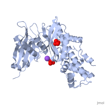
| - | | | + | <table><tr><td colspan='2'>[[1v4t]] is a 1 chain structure with sequence from [https://en.wikipedia.org/wiki/Homo_sapiens Homo sapiens]. Full crystallographic information is available from [http://oca.weizmann.ac.il/oca-bin/ocashort?id=1V4T OCA]. For a <b>guided tour on the structure components</b> use [https://proteopedia.org/fgij/fg.htm?mol=1V4T FirstGlance]. <br> |
| - | + | </td></tr><tr id='method'><td class="sblockLbl"><b>[[Empirical_models|Method:]]</b></td><td class="sblockDat" id="methodDat">X-ray diffraction, [[Resolution|Resolution]] 3.4Å</td></tr> | |
| - | + | <tr id='ligand'><td class="sblockLbl"><b>[[Ligand|Ligands:]]</b></td><td class="sblockDat" id="ligandDat"><scene name='pdbligand=NA:SODIUM+ION'>NA</scene>, <scene name='pdbligand=SO4:SULFATE+ION'>SO4</scene></td></tr> | |
| - | + | <tr id='resources'><td class="sblockLbl"><b>Resources:</b></td><td class="sblockDat"><span class='plainlinks'>[https://proteopedia.org/fgij/fg.htm?mol=1v4t FirstGlance], [http://oca.weizmann.ac.il/oca-bin/ocaids?id=1v4t OCA], [https://pdbe.org/1v4t PDBe], [https://www.rcsb.org/pdb/explore.do?structureId=1v4t RCSB], [https://www.ebi.ac.uk/pdbsum/1v4t PDBsum], [https://prosat.h-its.org/prosat/prosatexe?pdbcode=1v4t ProSAT]</span></td></tr> | |
| - | + | </table> | |
| - | + | == Disease == | |
| - | + | [https://www.uniprot.org/uniprot/HXK4_HUMAN HXK4_HUMAN] Defects in GCK are the cause of maturity-onset diabetes of the young type 2 (MODY2) [MIM:[https://omim.org/entry/125851 125851]; also shortened MODY-2. MODY is a form of diabetes that is characterized by an autosomal dominant mode of inheritance, onset in childhood or early adulthood (usually before 25 years of age), a primary defect in insulin secretion and frequent insulin-independence at the beginning of the disease.<ref>PMID:1502186</ref> <ref>PMID:1464666</ref> <ref>PMID:1303265</ref> <ref>PMID:8495817</ref> <ref>PMID:8325892</ref> <ref>PMID:8446612</ref> <ref>PMID:8168652</ref> <ref>PMID:9049484</ref> <ref>PMID:10694920</ref> <ref>PMID:9662401</ref> <ref>PMID:10588527</ref> <ref>PMID:11106831</ref> <ref>PMID:11372010</ref> Defects in GCK are the cause of familial hyperinsulinemic hypoglycemia type 3 (HHF3) [MIM:[https://omim.org/entry/602485 602485]; also known as persistent hyperinsulinemic hypoglycemia of infancy (PHHI) or congenital hyperinsulinism. HHF is the most common cause of persistent hypoglycemia in infancy. Unless early and aggressive intervention is undertaken, brain damage from recurrent episodes of hypoglycemia may occur.<ref>PMID:9435328</ref> | |
| + | == Function == | ||
| + | [https://www.uniprot.org/uniprot/HXK4_HUMAN HXK4_HUMAN] Catalyzes the initial step in utilization of glucose by the beta-cell and liver at physiological glucose concentration. Glucokinase has a high Km for glucose, and so it is effective only when glucose is abundant. The role of GCK is to provide G6P for the synthesis of glycogen. Pancreatic glucokinase plays an important role in modulating insulin secretion. Hepatic glucokinase helps to facilitate the uptake and conversion of glucose by acting as an insulin-sensitive determinant of hepatic glucose usage. | ||
| + | == Evolutionary Conservation == | ||
| + | [[Image:Consurf_key_small.gif|200px|right]] | ||
| + | Check<jmol> | ||
| + | <jmolCheckbox> | ||
| + | <scriptWhenChecked>; select protein; define ~consurf_to_do selected; consurf_initial_scene = true; script "/wiki/ConSurf/v4/1v4t_consurf.spt"</scriptWhenChecked> | ||
| + | <scriptWhenUnchecked>script /wiki/extensions/Proteopedia/spt/initialview01.spt</scriptWhenUnchecked> | ||
| + | <text>to colour the structure by Evolutionary Conservation</text> | ||
| + | </jmolCheckbox> | ||
| + | </jmol>, as determined by [http://consurfdb.tau.ac.il/ ConSurfDB]. You may read the [[Conservation%2C_Evolutionary|explanation]] of the method and the full data available from [http://bental.tau.ac.il/new_ConSurfDB/main_output.php?pdb_ID=1v4t ConSurf]. | ||
| + | <div style="clear:both"></div> | ||
| + | <div style="background-color:#fffaf0;"> | ||
| + | == Publication Abstract from PubMed == | ||
| + | Glucokinase is a monomeric enzyme that displays a low affinity for glucose and a sigmoidal saturation curve for its substrate, two properties that are important for its playing the role of a glucose sensor in pancreas and liver. The molecular basis for these two properties is not well understood. Herein we report the crystal structures of glucokinase in its active and inactive forms, which demonstrate that global conformational change, including domain reorganization, is induced by glucose binding. This suggests that the positive cooperativity of monomeric glucokinase obeys the "mnemonical mechanism" rather than the well-known concerted model. These structures also revealed an allosteric site through which small molecules may modulate the kinetic properties of the enzyme. This finding provided the mechanistic basis for activation of glucokinase as a potential therapeutic approach for treating type 2 diabetes mellitus. | ||
| - | + | Structural basis for allosteric regulation of the monomeric allosteric enzyme human glucokinase.,Kamata K, Mitsuya M, Nishimura T, Eiki J, Nagata Y Structure. 2004 Mar;12(3):429-38. PMID:15016359<ref>PMID:15016359</ref> | |
| + | From MEDLINE®/PubMed®, a database of the U.S. National Library of Medicine.<br> | ||
| + | </div> | ||
| + | <div class="pdbe-citations 1v4t" style="background-color:#fffaf0;"></div> | ||
| - | == | + | ==See Also== |
| - | + | *[[Hexokinase 3D structures|Hexokinase 3D structures]] | |
| - | + | == References == | |
| - | == | + | <references/> |
| - | + | __TOC__ | |
| - | + | </StructureSection> | |
| - | + | ||
| - | + | ||
| - | + | ||
[[Category: Homo sapiens]] | [[Category: Homo sapiens]] | ||
| - | [[Category: | + | [[Category: Large Structures]] |
| - | [[Category: Eiki | + | [[Category: Eiki J]] |
| - | [[Category: Kamata | + | [[Category: Kamata K]] |
| - | [[Category: Mitsuya | + | [[Category: Mitsuya M]] |
| - | [[Category: Nagata | + | [[Category: Nagata Y]] |
| - | [[Category: Nishimura | + | [[Category: Nishimura T]] |
| - | + | ||
| - | + | ||
| - | + | ||
| - | + | ||
| - | + | ||
Current revision
Crystal structure of human glucokinase
| |||||||||||

Categories: Homo sapiens | Large Structures | Eiki J | Kamata K | Mitsuya M | Nagata Y | Nishimura T
