This old version of Proteopedia is provided for student assignments while the new version is undergoing repairs. Content and edits done in this old version of Proteopedia after March 1, 2026 will eventually be lost when it is retired in about June of 2026.
Apply for new accounts at the new Proteopedia. Your logins will work in both the old and new versions.
1pg1
From Proteopedia
(Difference between revisions)
(New page: 200px <!-- The line below this paragraph, containing "STRUCTURE_1pg1", creates the "Structure Box" on the page. You may change the PDB parameter (which sets the PD...) |
|||
| (10 intermediate revisions not shown.) | |||
| Line 1: | Line 1: | ||
| - | [[Image:1pg1.png|left|200px]] | ||
| - | < | + | ==PROTEGRIN 1 (PG1) FROM PORCINE LEUKOCYTES, NMR, 20 STRUCTURES== |
| - | + | <StructureSection load='1pg1' size='340' side='right'caption='[[1pg1]]' scene=''> | |
| - | + | == Structural highlights == | |
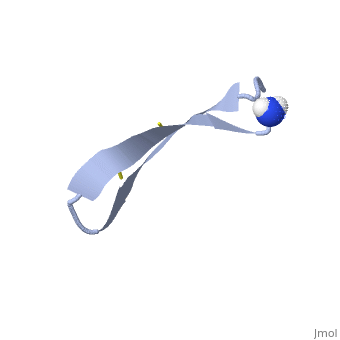
| - | + | <table><tr><td colspan='2'>[[1pg1]] is a 1 chain structure with sequence from [https://en.wikipedia.org/wiki/Sus_scrofa Sus scrofa]. Full experimental information is available from [http://oca.weizmann.ac.il/oca-bin/ocashort?id=1PG1 OCA]. For a <b>guided tour on the structure components</b> use [https://proteopedia.org/fgij/fg.htm?mol=1PG1 FirstGlance]. <br> | |
| - | or | + | </td></tr><tr id='method'><td class="sblockLbl"><b>[[Empirical_models|Method:]]</b></td><td class="sblockDat" id="methodDat">Solution NMR</td></tr> |
| - | -- | + | <tr id='ligand'><td class="sblockLbl"><b>[[Ligand|Ligands:]]</b></td><td class="sblockDat" id="ligandDat"><scene name='pdbligand=NH2:AMINO+GROUP'>NH2</scene></td></tr> |
| - | + | <tr id='resources'><td class="sblockLbl"><b>Resources:</b></td><td class="sblockDat"><span class='plainlinks'>[https://proteopedia.org/fgij/fg.htm?mol=1pg1 FirstGlance], [http://oca.weizmann.ac.il/oca-bin/ocaids?id=1pg1 OCA], [https://pdbe.org/1pg1 PDBe], [https://www.rcsb.org/pdb/explore.do?structureId=1pg1 RCSB], [https://www.ebi.ac.uk/pdbsum/1pg1 PDBsum], [https://prosat.h-its.org/prosat/prosatexe?pdbcode=1pg1 ProSAT]</span></td></tr> | |
| + | </table> | ||
| + | == Function == | ||
| + | [https://www.uniprot.org/uniprot/PG1_PIG PG1_PIG] Microbicidal activity. Active against E.coli, Listeria monocytogenes and C.albicans, in vitro. | ||
| + | <div style="background-color:#fffaf0;"> | ||
| + | == Publication Abstract from PubMed == | ||
| + | BACKGROUND: The protegrins are a family of arginine- and cysteine-rich cationic peptides found in porcine leukocytes that exhibit a broad range of antimicrobial and antiviral activities. They are composed of 16-18 amino-acid residues including four cysteines, which form two disulfide linkages. To begin to understand the mechanism of action of these peptides, we set out to determine the structure of protegrin-1 (PG-1). RESULTS: We used two-dimensional homonuclear nuclear magnetic resonance spectroscopy to study the conformation of both natural and synthetic PG-1 under several conditions. A refined three-dimensional structure of synthetic PG-1 is presented. CONCLUSIONS: Both synthetic and natural protegrin-1 form a well-defined structure in solution composed primarily of a two-stranded antiparallel beta sheet, with strands connected by a beta turn. The structure of PG-1 suggests ways in which the peptide may interact with itself or other molecules to form the membrane pores and the large membrane-associated assemblages observed in protegrin-treated, gram-negative bacteria. | ||
| - | + | Solution structure of protegrin-1, a broad-spectrum antimicrobial peptide from porcine leukocytes.,Fahrner RL, Dieckmann T, Harwig SS, Lehrer RI, Eisenberg D, Feigon J Chem Biol. 1996 Jul;3(7):543-50. PMID:8807886<ref>PMID:8807886</ref> | |
| + | From MEDLINE®/PubMed®, a database of the U.S. National Library of Medicine.<br> | ||
| + | </div> | ||
| + | <div class="pdbe-citations 1pg1" style="background-color:#fffaf0;"></div> | ||
| - | + | ==See Also== | |
| - | + | *[[Protegrin|Protegrin]] | |
| - | + | == References == | |
| - | + | <references/> | |
| - | + | __TOC__ | |
| - | + | </StructureSection> | |
| - | == | + | [[Category: Large Structures]] |
| - | [[ | + | |
| - | + | ||
| - | == | + | |
| - | < | + | |
[[Category: Sus scrofa]] | [[Category: Sus scrofa]] | ||
| - | [[Category: Dieckmann | + | [[Category: Dieckmann T]] |
| - | [[Category: Eisenberg | + | [[Category: Eisenberg D]] |
| - | [[Category: Fahrner | + | [[Category: Fahrner RL]] |
| - | [[Category: Feigon | + | [[Category: Feigon J]] |
| - | [[Category: Harwig | + | [[Category: Harwig SSL]] |
| - | [[Category: Lehrer | + | [[Category: Lehrer RI]] |
| - | + | ||
Current revision
PROTEGRIN 1 (PG1) FROM PORCINE LEUKOCYTES, NMR, 20 STRUCTURES
| |||||||||||
