1cii
From Proteopedia
(Difference between revisions)
| (13 intermediate revisions not shown.) | |||
| Line 1: | Line 1: | ||
| - | [[Image:1cii.gif|left|200px]] | ||
| - | + | ==COLICIN IA== | |
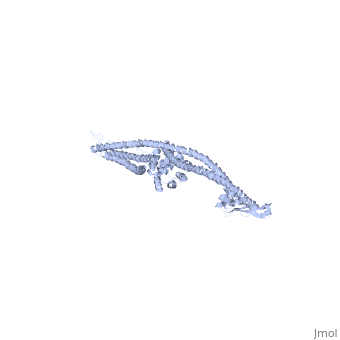
| - | + | <StructureSection load='1cii' size='340' side='right'caption='[[1cii]], [[Resolution|resolution]] 3.00Å' scene=''> | |
| - | + | == Structural highlights == | |
| - | + | <table><tr><td colspan='2'>[[1cii]] is a 1 chain structure with sequence from [https://en.wikipedia.org/wiki/Escherichia_coli Escherichia coli]. Full crystallographic information is available from [http://oca.weizmann.ac.il/oca-bin/ocashort?id=1CII OCA]. For a <b>guided tour on the structure components</b> use [https://proteopedia.org/fgij/fg.htm?mol=1CII FirstGlance]. <br> | |
| - | + | </td></tr><tr id='method'><td class="sblockLbl"><b>[[Empirical_models|Method:]]</b></td><td class="sblockDat" id="methodDat">X-ray diffraction, [[Resolution|Resolution]] 3Å</td></tr> | |
| - | + | <tr id='resources'><td class="sblockLbl"><b>Resources:</b></td><td class="sblockDat"><span class='plainlinks'>[https://proteopedia.org/fgij/fg.htm?mol=1cii FirstGlance], [http://oca.weizmann.ac.il/oca-bin/ocaids?id=1cii OCA], [https://pdbe.org/1cii PDBe], [https://www.rcsb.org/pdb/explore.do?structureId=1cii RCSB], [https://www.ebi.ac.uk/pdbsum/1cii PDBsum], [https://prosat.h-its.org/prosat/prosatexe?pdbcode=1cii ProSAT]</span></td></tr> | |
| - | + | </table> | |
| - | + | == Function == | |
| - | + | [https://www.uniprot.org/uniprot/CEIA_ECOLX CEIA_ECOLX] This colicin is a channel-forming colicin. This class of transmembrane toxins depolarize the cytoplasmic membrane, leading to dissipation of cellular energy. Colicins are polypeptide toxins produced by and active against E.coli and closely related bacteria. | |
| - | + | == Evolutionary Conservation == | |
| + | [[Image:Consurf_key_small.gif|200px|right]] | ||
| + | Check<jmol> | ||
| + | <jmolCheckbox> | ||
| + | <scriptWhenChecked>; select protein; define ~consurf_to_do selected; consurf_initial_scene = true; script "/wiki/ConSurf/ci/1cii_consurf.spt"</scriptWhenChecked> | ||
| + | <scriptWhenUnchecked>script /wiki/extensions/Proteopedia/spt/initialview01.spt</scriptWhenUnchecked> | ||
| + | <text>to colour the structure by Evolutionary Conservation</text> | ||
| + | </jmolCheckbox> | ||
| + | </jmol>, as determined by [http://consurfdb.tau.ac.il/ ConSurfDB]. You may read the [[Conservation%2C_Evolutionary|explanation]] of the method and the full data available from [http://bental.tau.ac.il/new_ConSurfDB/main_output.php?pdb_ID=1cii ConSurf]. | ||
| + | <div style="clear:both"></div> | ||
| - | + | ==See Also== | |
| - | + | *[[Colicin|Colicin]] | |
| - | + | *[[Colicin 3D structures|Colicin 3D structures]] | |
| - | == | + | __TOC__ |
| - | + | </StructureSection> | |
| - | + | ||
| - | + | ||
| - | + | ||
| - | + | ||
| - | + | ||
| - | + | ||
[[Category: Escherichia coli]] | [[Category: Escherichia coli]] | ||
| - | [[Category: | + | [[Category: Large Structures]] |
| - | [[Category: Freymann | + | [[Category: Freymann D]] |
| - | [[Category: Ghosh | + | [[Category: Ghosh P]] |
| - | [[Category: Stroud | + | [[Category: Stroud R]] |
| - | [[Category: Wiener | + | [[Category: Wiener M]] |
| - | + | ||
| - | + | ||
| - | + | ||
| - | + | ||
| - | + | ||
| - | + | ||
Current revision
COLICIN IA
| |||||||||||