We apologize for Proteopedia being slow to respond. For the past two years, a new implementation of Proteopedia has been being built. Soon, it will replace this 18-year old system. All existing content will be moved to the new system at a date that will be announced here.
1xbn
From Proteopedia
(Difference between revisions)
| Line 3: | Line 3: | ||
<StructureSection load='1xbn' size='340' side='right'caption='[[1xbn]], [[Resolution|resolution]] 2.50Å' scene=''> | <StructureSection load='1xbn' size='340' side='right'caption='[[1xbn]], [[Resolution|resolution]] 2.50Å' scene=''> | ||
== Structural highlights == | == Structural highlights == | ||
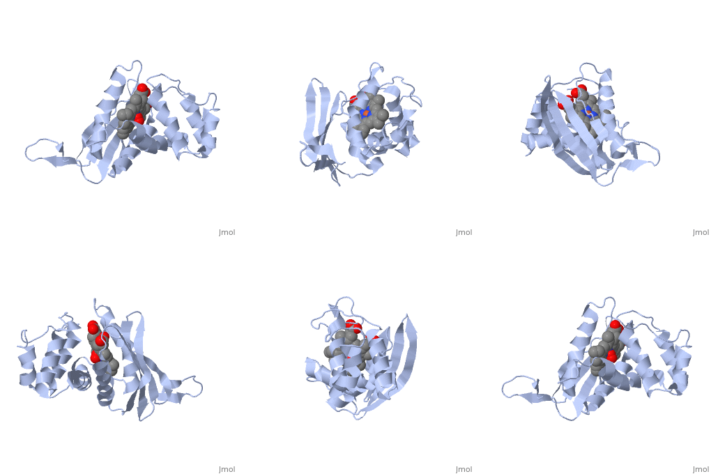
| - | <table><tr><td colspan='2'>[[1xbn]] is a 1 chain structure with sequence from [ | + | <table><tr><td colspan='2'>[[1xbn]] is a 1 chain structure with sequence from [https://en.wikipedia.org/wiki/Caldanaerobacter_subterraneus_subsp._tengcongensis_MB4 Caldanaerobacter subterraneus subsp. tengcongensis MB4]. Full crystallographic information is available from [http://oca.weizmann.ac.il/oca-bin/ocashort?id=1XBN OCA]. For a <b>guided tour on the structure components</b> use [https://proteopedia.org/fgij/fg.htm?mol=1XBN FirstGlance]. <br> |
| - | </td></tr><tr id=' | + | </td></tr><tr id='method'><td class="sblockLbl"><b>[[Empirical_models|Method:]]</b></td><td class="sblockDat" id="methodDat">X-ray diffraction, [[Resolution|Resolution]] 2.5Å</td></tr> |
| - | <tr id=' | + | <tr id='ligand'><td class="sblockLbl"><b>[[Ligand|Ligands:]]</b></td><td class="sblockDat" id="ligandDat"><scene name='pdbligand=HEM:PROTOPORPHYRIN+IX+CONTAINING+FE'>HEM</scene>, <scene name='pdbligand=OXY:OXYGEN+MOLECULE'>OXY</scene></td></tr> |
| - | <tr id='resources'><td class="sblockLbl"><b>Resources:</b></td><td class="sblockDat"><span class='plainlinks'>[ | + | <tr id='resources'><td class="sblockLbl"><b>Resources:</b></td><td class="sblockDat"><span class='plainlinks'>[https://proteopedia.org/fgij/fg.htm?mol=1xbn FirstGlance], [http://oca.weizmann.ac.il/oca-bin/ocaids?id=1xbn OCA], [https://pdbe.org/1xbn PDBe], [https://www.rcsb.org/pdb/explore.do?structureId=1xbn RCSB], [https://www.ebi.ac.uk/pdbsum/1xbn PDBsum], [https://prosat.h-its.org/prosat/prosatexe?pdbcode=1xbn ProSAT]</span></td></tr> |
</table> | </table> | ||
| + | == Function == | ||
| + | [https://www.uniprot.org/uniprot/Q8RBX6_CALS4 Q8RBX6_CALS4] | ||
== Evolutionary Conservation == | == Evolutionary Conservation == | ||
[[Image:Consurf_key_small.gif|200px|right]] | [[Image:Consurf_key_small.gif|200px|right]] | ||
| Line 18: | Line 20: | ||
</jmol>, as determined by [http://consurfdb.tau.ac.il/ ConSurfDB]. You may read the [[Conservation%2C_Evolutionary|explanation]] of the method and the full data available from [http://bental.tau.ac.il/new_ConSurfDB/main_output.php?pdb_ID=1xbn ConSurf]. | </jmol>, as determined by [http://consurfdb.tau.ac.il/ ConSurfDB]. You may read the [[Conservation%2C_Evolutionary|explanation]] of the method and the full data available from [http://bental.tau.ac.il/new_ConSurfDB/main_output.php?pdb_ID=1xbn ConSurf]. | ||
<div style="clear:both"></div> | <div style="clear:both"></div> | ||
| - | <div style="background-color:#fffaf0;"> | ||
| - | == Publication Abstract from PubMed == | ||
| - | Nitric oxide (NO) is extremely toxic to Clostridium botulinum, but its molecular targets are unknown. Here, we identify a heme protein sensor (SONO) that displays femtomolar affinity for NO. The crystal structure of the SONO heme domain reveals a previously undescribed fold and a strategically placed tyrosine residue that modulates heme-nitrosyl coordination. Furthermore, the domain architecture of a SONO ortholog cloned from Chlamydomonas reinhardtii indicates that NO signaling through cyclic guanosine monophosphate arose before the origin of multicellular eukaryotes. Our findings have broad implications for understanding bacterial responses to NO, as well as for the activation of mammalian NO-sensitive guanylyl cyclase. | ||
| - | |||
| - | Femtomolar sensitivity of a NO sensor from Clostridium botulinum.,Nioche P, Berka V, Vipond J, Minton N, Tsai AL, Raman CS Science. 2004 Nov 26;306(5701):1550-3. Epub 2004 Oct 7. PMID:15472039<ref>PMID:15472039</ref> | ||
| - | |||
| - | From MEDLINE®/PubMed®, a database of the U.S. National Library of Medicine.<br> | ||
| - | </div> | ||
| - | <div class="pdbe-citations 1xbn" style="background-color:#fffaf0;"></div> | ||
==See Also== | ==See Also== | ||
*[[Chemotaxis protein 3D structures|Chemotaxis protein 3D structures]] | *[[Chemotaxis protein 3D structures|Chemotaxis protein 3D structures]] | ||
*[[Methyl-accepting chemotaxis protein|Methyl-accepting chemotaxis protein]] | *[[Methyl-accepting chemotaxis protein|Methyl-accepting chemotaxis protein]] | ||
| - | == References == | ||
| - | <references/> | ||
__TOC__ | __TOC__ | ||
</StructureSection> | </StructureSection> | ||
| - | [[Category: | + | [[Category: Caldanaerobacter subterraneus subsp. tengcongensis MB4]] |
[[Category: Large Structures]] | [[Category: Large Structures]] | ||
| - | [[Category: Nioche | + | [[Category: Nioche P]] |
| - | [[Category: Raman | + | [[Category: Raman CS]] |
| - | + | ||
| - | + | ||
| - | + | ||
| - | + | ||
| - | + | ||
Current revision
Crystal structure of a bacterial nitric oxide sensor: an ortholog of mammalian soluble guanylate cyclase heme domain
| |||||||||||


